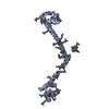
 Movie
Movie Controller


+ Open data

- Basic information
| Entry | Database: PDB / ID: 2nzi | ||||||
|---|---|---|---|---|---|---|---|
| Title | Crystal structure of domains A168-A170 from titin | ||||||
 Components Components | Titin | ||||||
 Keywords Keywords | TRANSFERASE / Ig-domain / FnIII-domain | ||||||
| Function / homology |  Function and homology information Function and homology informationsarcomerogenesis / structural molecule activity conferring elasticity / telethonin binding / skeletal muscle myosin thick filament assembly / cardiac myofibril assembly / muscle alpha-actinin binding / detection of muscle stretch / cardiac muscle tissue morphogenesis / regulation of catalytic activity / cardiac muscle hypertrophy ...sarcomerogenesis / structural molecule activity conferring elasticity / telethonin binding / skeletal muscle myosin thick filament assembly / cardiac myofibril assembly / muscle alpha-actinin binding / detection of muscle stretch / cardiac muscle tissue morphogenesis / regulation of catalytic activity / cardiac muscle hypertrophy / mitotic chromosome condensation / Striated Muscle Contraction / M band / actinin binding / I band / cardiac muscle cell development / regulation of protein kinase activity / sarcomere organization / structural constituent of muscle / skeletal muscle thin filament assembly / striated muscle thin filament / striated muscle contraction / protein kinase A signaling / cardiac muscle contraction / muscle contraction / condensed nuclear chromosome / positive regulation of protein secretion / Z disc / response to calcium ion / : / actin filament binding / Platelet degranulation / protein tyrosine kinase activity / protease binding / calmodulin binding / non-specific serine/threonine protein kinase / phosphorylation / protein serine kinase activity / protein serine/threonine kinase activity / calcium ion binding / positive regulation of gene expression / protein kinase binding / enzyme binding / extracellular exosome / extracellular region / ATP binding / identical protein binding / cytosolSimilarity search - Function | ||||||
| Biological species |  Homo sapiens (human) Homo sapiens (human) | ||||||
| Method | X-RAY DIFFRACTION / SYNCHROTRON / MAD / Resolution: 2.9 Å | ||||||
 Authors Authors | Mrosek, M.C. / Labeit, D. / Labeit, S. / Mayans, O. | ||||||
 Citation Citation | Journal: Faseb J. / Year: 2007 Title: Molecular determinants for the recruitment of the ubiquitin-ligase MuRF-1 onto M-line titin. Authors: Mrosek, M. / Labeit, D. / Witt, S. / Heerklotz, H. / von Castelmur, E. / Labeit, S. / Mayans, O. | ||||||
| History |
|
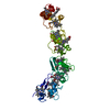
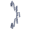
- Structure visualization
| Structure viewer | Molecule: MolmilJmol/JSmol |
|---|
- Downloads & links
-Download
| PDBx/mmCIF format | 2nzi.cif.gz | 113.8 KB | Display | PDBx/mmCIF format |
|---|---|---|---|---|
| PDB format | pdb2nzi.ent.gz | 90.6 KB | Display | PDB format |
| PDBx/mmJSON format | 2nzi.json.gz | Tree view | PDBx/mmJSON format | |
| Others |  Other downloads Other downloads |
-Validation report
| Arichive directory | https://data.pdbj.org/pub/pdb/validation_reports/nz/2nziftp://data.pdbj.org/pub/pdb/validation_reports/nz/2nzi | HTTPS FTP |
|---|
-Related structure data
| Similar structure data |
|---|
-Links
 PDBj
PDBj


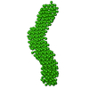
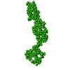
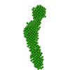
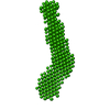
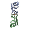
- Assembly
| Deposited unit | 
| ||||||||
|---|---|---|---|---|---|---|---|---|---|
| 1 | 
| ||||||||
| 2 | 
| ||||||||
| Unit cell |
| ||||||||
| Details | The asymmetric unit contains two biological units. Chain A and Chain B are a biological unit each. |
-Components
| #1: Protein | / Connectin / Rhabdomyosarcoma antigen MU-RMS-40.14 Mass: 33920.359 Da / Num. of mol.: 2 / Fragment: Residues 31854-32155 Source method: isolated from a genetically manipulated source Source: (gene. exp.) Homo sapiens (human) / Gene: TTN / Plasmid: pETM-11 / Production host:  Escherichia coli (E. coli) / Strain (production host): BL21(DE3)Rosetta Escherichia coli (E. coli) / Strain (production host): BL21(DE3)RosettaReferences: UniProt: Q8WZ42, non-specific serine/threonine protein kinase#2: Water | ChemComp-HOH / | Water Mass: 18.015 Da / Num. of mol.: 44 / Source method: isolated from a natural source / Formula: H2O Mass: 18.015 Da / Num. of mol.: 44 / Source method: isolated from a natural source / Formula: H2O |
|---|
-Experimental details
-Experiment
| Experiment | Method: X-RAY DIFFRACTION / Number of used crystals: 1 |
|---|
- Sample preparation
| Crystal | Density Matthews: 4.52 Å3/Da / Density % sol: 72.78 % |
|---|---|
| Crystal grow | Temperature: 298 K / Method: vapor diffusion, hanging drop / pH: 5.5 Details: 2.1M NaCl, 0.1M MES, 0.6M Li2SO4, pH 5.5, VAPOR DIFFUSION, HANGING DROP, temperature 298K |
-Data collection
| Diffraction | Mean temperature: 100 K |
|---|---|
| Diffraction source | Source: SYNCHROTRON / Site: ESRF  / Beamline: ID29 / Wavelength: 0.9792 Å / Beamline: ID29 / Wavelength: 0.9792 Å |
| Detector | Type: ADSC QUANTUM 210 / Detector: CCD / Date: Nov 3, 2004 |
| Radiation | Protocol: MAD / Monochromatic (M) / Laue (L): M / Scattering type: x-ray |
| Radiation wavelength | Wavelength: 0.9792 Å / Relative weight: 1 |
| Reflection | Resolution: 2.9→15 Å / Num. all: 27728 / Num. obs: 26792 / % possible obs: 96.6 % / Redundancy: 6.3 % / Rsym value: 0.075 / Net I/σ(I): 22.7 |
| Reflection shell | Resolution: 2.9→3 Å / Redundancy: 6.3 % / Mean I/σ(I) obs: 3.6 / Num. unique all: 2627 / Rsym value: 0.531 / % possible all: 98.6 |
- Processing
| Software |
| |||||||||||||||||||||||||
|---|---|---|---|---|---|---|---|---|---|---|---|---|---|---|---|---|---|---|---|---|---|---|---|---|---|---|
| Refinement | Method to determine structure: MAD / Resolution: 2.9→15 Å / Isotropic thermal model: Isotropic / Cross valid method: THROUGHOUT / σ(F): 0 / Stereochemistry target values: Engh & Huber
| |||||||||||||||||||||||||
| Displacement parameters | Biso mean: 38.66 Å2
| |||||||||||||||||||||||||
| Refinement step | Cycle: LAST / Resolution: 2.9→15 Å
|
