Mass: 18.015 Da / Num. of mol.: 34 / Source method: isolated from a natural source / Formula: H2O
Sequence details
THE CONSTRUCT WAS EXPRESSED WITH A PURIFICATION TAG MGSDKIHHHHHHENLYFQG. THE TAG WAS REMOVED WITH ...THE CONSTRUCT WAS EXPRESSED WITH A PURIFICATION TAG MGSDKIHHHHHHENLYFQG. THE TAG WAS REMOVED WITH TEV PROTEASE LEAVING ONLY A GLYCINE (0) FOLLOWED BY THE TARGET SEQUENCE.
-
Experimental details
-
Experiment
Experiment
Method: X-RAY DIFFRACTION / Number of used crystals: 1
-
Sample preparation
Crystal
Density Matthews: 2.47 Å3/Da / Density % sol: 50.28 %
Crystal grow
Temperature: 293 K / Method: vapor diffusion, sitting drop Details: 20.0000% polyethylene glycol 3350, 0.2500M lithium sulfate, NANODROP, VAPOR DIFFUSION, SITTING DROP, temperature 293K
Resolution: 2.81→46.932 Å / Num. obs: 28370 / % possible obs: 98.5 % / Observed criterion σ(I): -3 / Biso Wilson estimate: 63.565 Å2 / Rmerge(I) obs: 0.124 / Net I/σ(I): 8.29
Reflection shell
Resolution (Å)
Rmerge(I) obs
Mean I/σ(I) obs
Num. measured obs
Num. unique obs
Diffraction-ID
% possible all
2.81-2.91
0.757
2.3
8828
2581
1
91
2.91-3.03
0.684
2.4
9986
2850
1
99
3.03-3.16
0.495
3.3
9382
2667
1
99.5
3.16-3.33
0.333
4.6
10377
2887
1
99.6
3.33-3.54
0.227
6.2
10265
2852
1
99.6
3.54-3.81
0.149
8.4
10239
2809
1
99.9
3.81-4.19
0.103
10.7
10396
2849
1
99.8
4.19-4.79
0.078
12.7
10537
2893
1
99.8
4.79-6
0.079
12.7
10434
2884
1
99.5
6-46.932
0.046
17.9
10772
3097
1
97.5
-
Phasing
Phasing
Method: MAD
-
Processing
Software
Name
Version
Classification
NB
BUSTER
2.8.0
refinement
PHENIX
refinement
SHELX
phasing
XSCALE
datascaling
PDB_EXTRACT
3.006
dataextraction
XDS
datareduction
SHELXD
phasing
autoSHARP
phasing
MolProbity
3beta29
modelbuilding
Refinement
Method to determine structure: MAD / Resolution: 2.81→46.932 Å / Cor.coef. Fo:Fc: 0.918 / Cor.coef. Fo:Fc free: 0.879 / Occupancy max: 1 / Occupancy min: 0.5 / Cross valid method: THROUGHOUT / σ(F): 0 Details: 1. A MET-INHIBITION PROTOCOL WAS USED FOR SELENOMETHIONINE INCORPORATION DURING PROTEIN EXPRESSION. THE OCCUPANCY OF THE SE ATOMS IN THE MSE RESIDUES WAS REDUCED TO 0.75 FOR THE REDUCED ...Details: 1. A MET-INHIBITION PROTOCOL WAS USED FOR SELENOMETHIONINE INCORPORATION DURING PROTEIN EXPRESSION. THE OCCUPANCY OF THE SE ATOMS IN THE MSE RESIDUES WAS REDUCED TO 0.75 FOR THE REDUCED SCATTERING POWER DUE TO PARTIAL S-MET INCORPORATION. 2. SULFATE MODELED ARE PRESENT IN CRYSTLLIZATION CONDITIONS. 3. RAMACHANDRAN OUTLIER (A6) IS LOCATED IN REGION OF POOR DENSITY.
In the structure databanks used in Yorodumi, some data are registered as the other names, "COVID-19 virus" and "2019-nCoV". Here are the details of the virus and the list of structure data.
Jan 31, 2019. EMDB accession codes are about to change! (news from PDBe EMDB page)
EMDB accession codes are about to change! (news from PDBe EMDB page)
The allocation of 4 digits for EMDB accession codes will soon come to an end. Whilst these codes will remain in use, new EMDB accession codes will include an additional digit and will expand incrementally as the available range of codes is exhausted. The current 4-digit format prefixed with “EMD-” (i.e. EMD-XXXX) will advance to a 5-digit format (i.e. EMD-XXXXX), and so on. It is currently estimated that the 4-digit codes will be depleted around Spring 2019, at which point the 5-digit format will come into force.
The EM Navigator/Yorodumi systems omit the EMD- prefix.
Related info.:Q: What is EMD? / ID/Accession-code notation in Yorodumi/EM Navigator
Yorodumi is a browser for structure data from EMDB, PDB, SASBDB, etc.
This page is also the successor to EM Navigator detail page, and also detail information page/front-end page for Omokage search.
The word "yorodu" (or yorozu) is an old Japanese word meaning "ten thousand". "mi" (miru) is to see.
Related info.:EMDB / PDB / SASBDB / Comparison of 3 databanks / Yorodumi Search / Aug 31, 2016. New EM Navigator & Yorodumi / Yorodumi Papers / Jmol/JSmol / Function and homology information / Changes in new EM Navigator and Yorodumi
 Movie
Movie Controller

 Yorodumi
 Open data
 Basic information
 Components
 Transcriptional regulation
  Keywords
 Function and homology information
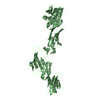
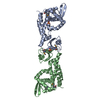
 Jannaschia sp. (bacteria)
 Authors
 Citation
 Structure visualization
 Downloads & links
 Other downloads


 PDBj
PDBj Assembly


 Mass: 96.063 Da / Num. of mol.: 10 / Source method: obtained synthetically / Formula: SO4
 Mass: 18.015 Da / Num. of mol.: 34 / Source method: isolated from a natural source / Formula: H2O
 Sample preparation
 / Beamline: BL9-2 / Wavelength: 0.97939,0.91162,0.97925
 Processing