 Movie
Movie Controller


+ Open data

- Basic information
| Entry | Database: PDB / ID: 3b43 | ||||||
|---|---|---|---|---|---|---|---|
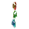
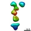
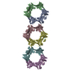
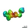
| Title | I-band fragment I65-I70 from titin | ||||||
 Components Components | Titin | ||||||
 Keywords Keywords | STRUCTURAL PROTEIN / I-set Ig fold / extended poly-Ig filament / titin / elastic filament | ||||||
| Function / homology |  Function and homology information Function and homology information | ||||||
| Biological species |  Oryctolagus cuniculus (rabbit) Oryctolagus cuniculus (rabbit) | ||||||
| Method | X-RAY DIFFRACTION / SYNCHROTRON / MIRAS / Resolution: 3.3 Å | ||||||
 Authors Authors | von Castelmur, E. / Marino, M. / Labeit, D. / Labeit, S. / Mayans, O. | ||||||
 Citation Citation | Journal: Proc.Natl.Acad.Sci.Usa / Year: 2008 Title: A regular pattern of Ig super-motifs defines segmental flexibility as the elastic mechanism of the titin chain Authors: von Castelmur, E. / Marino, M. / Svergun, D.I. / Kreplak, L. / Ucurum-Fotiadis, Z. / Konarev, P.V. / Urzhumtsev, A. / Labeit, D. / Labeit, S. / Mayans, O. #1: Journal: J.Muscle Res.Cell.Motil. / Year: 2005 Title: Poly-Ig tandems from I-band titin share extended domain arrangements irrespective of the distinct features of their modular constituents Authors: Marino, M. / Svergun, D.I. / Kreplak, L. / Konarev, P.V. / Maco, B. / Labeit, D. / Mayans, O. | ||||||
| History |
|
- Structure visualization
| Structure viewer | Molecule: MolmilJmol/JSmol |
|---|
- Downloads & links
-Download
| PDBx/mmCIF format | 3b43.cif.gz | 233.4 KB | Display | PDBx/mmCIF format |
|---|---|---|---|---|
| PDB format | pdb3b43.ent.gz | 198.2 KB | Display | PDB format |
| PDBx/mmJSON format | 3b43.json.gz | Tree view | PDBx/mmJSON format | |
| Others |  Other downloads Other downloads |
-Validation report
| Arichive directory | https://data.pdbj.org/pub/pdb/validation_reports/b4/3b43ftp://data.pdbj.org/pub/pdb/validation_reports/b4/3b43 | HTTPS FTP |
|---|
-Related structure data

-Links
 PDBj
PDBj

- Assembly
| Deposited unit | 
| ||||||||
|---|---|---|---|---|---|---|---|---|---|
| 1 |
| ||||||||
| Unit cell |
|
-Components
| #1: Protein | Mass: 62888.961 Da / Num. of mol.: 1 / Fragment: I65-I70 Source method: isolated from a genetically manipulated source Source: (gene. exp.) Oryctolagus cuniculus (rabbit) / Plasmid: pETM-11 / Production host:  Escherichia coli (E. coli) / Strain (production host): BL21 (DE3) Rosetta Escherichia coli (E. coli) / Strain (production host): BL21 (DE3) RosettaReferences: UniProt: D0VWS0*PLUS, non-specific serine/threonine protein kinase |
|---|---|
| Sequence details | THE DATABASE SEQUENCE ENTRY COVERING THIS CONSTRUCT IS EMBL Y14852, RABBIT SOLEUS TITIN MRNA, BASES ...THE DATABASE SEQUENCE ENTRY COVERING THIS CONSTRUCT IS EMBL Y14852, RABBIT SOLEUS TITIN MRNA, BASES 11881-13578. THE CLOSEST HOMOLOG IS HUMAN TITIN SWISSPROT ENTRY Q8WZ42, RESIDUES 7946-8511. |
-Experimental details
-Experiment
| Experiment | Method: X-RAY DIFFRACTION / Number of used crystals: 1 |
|---|
- Sample preparation
| Crystal | Density Matthews: 3.81 Å3/Da / Density % sol: 67.72 % |
|---|---|
| Crystal grow | Temperature: 298 K / Method: vapor diffusion, hanging drop / pH: 4.9 Details: pH 4.9, VAPOR DIFFUSION, HANGING DROP, temperature 298K |
-Data collection
| Diffraction | Mean temperature: 100 K | |||||||||||||||||||||||||||||||||||||||||||||||||||||||||||||||||||||||||||||||||||||||||||
|---|---|---|---|---|---|---|---|---|---|---|---|---|---|---|---|---|---|---|---|---|---|---|---|---|---|---|---|---|---|---|---|---|---|---|---|---|---|---|---|---|---|---|---|---|---|---|---|---|---|---|---|---|---|---|---|---|---|---|---|---|---|---|---|---|---|---|---|---|---|---|---|---|---|---|---|---|---|---|---|---|---|---|---|---|---|---|---|---|---|---|---|---|
| Diffraction source | Source: SYNCHROTRON / Site: ESRF  / Beamline: ID29 / Wavelength: 0.9792 Å / Beamline: ID29 / Wavelength: 0.9792 Å | |||||||||||||||||||||||||||||||||||||||||||||||||||||||||||||||||||||||||||||||||||||||||||
| Detector | Type: ADSC QUANTUM 210 / Detector: CCD / Date: Nov 3, 2004 | |||||||||||||||||||||||||||||||||||||||||||||||||||||||||||||||||||||||||||||||||||||||||||
| Radiation | Protocol: SINGLE WAVELENGTH / Monochromatic (M) / Laue (L): M / Scattering type: x-ray | |||||||||||||||||||||||||||||||||||||||||||||||||||||||||||||||||||||||||||||||||||||||||||
| Radiation wavelength | Wavelength: 0.9792 Å / Relative weight: 1 | |||||||||||||||||||||||||||||||||||||||||||||||||||||||||||||||||||||||||||||||||||||||||||
| Reflection | Resolution: 3.3→17 Å / Num. obs: 14953 / % possible obs: 98.57 % / Observed criterion σ(I): -3 / Biso Wilson estimate: 82.5 Å2 / Rmerge(I) obs: 0.08 / Net I/σ(I): 19.6 | |||||||||||||||||||||||||||||||||||||||||||||||||||||||||||||||||||||||||||||||||||||||||||
| Reflection shell | Diffraction-ID: 1
|
-Phasing
| Phasing | Method: MIRAS |
|---|
- Processing
| Software |
| |||||||||||||||||||||||||||||||||||||||||||||||||||||||||||||||||||||||||||||||||||||||||||||||||||||||||||||||||||||||||||||||||||||||||||||||||||||||||||||||||||||||||||||||
|---|---|---|---|---|---|---|---|---|---|---|---|---|---|---|---|---|---|---|---|---|---|---|---|---|---|---|---|---|---|---|---|---|---|---|---|---|---|---|---|---|---|---|---|---|---|---|---|---|---|---|---|---|---|---|---|---|---|---|---|---|---|---|---|---|---|---|---|---|---|---|---|---|---|---|---|---|---|---|---|---|---|---|---|---|---|---|---|---|---|---|---|---|---|---|---|---|---|---|---|---|---|---|---|---|---|---|---|---|---|---|---|---|---|---|---|---|---|---|---|---|---|---|---|---|---|---|---|---|---|---|---|---|---|---|---|---|---|---|---|---|---|---|---|---|---|---|---|---|---|---|---|---|---|---|---|---|---|---|---|---|---|---|---|---|---|---|---|---|---|---|---|---|---|---|---|---|
| Refinement | Method to determine structure: MIRAS / Resolution: 3.3→16.985 Å / FOM work R set: 0.794 / SU ML: 0.46 / Phase error: 26.76 / Stereochemistry target values: ML / Details: This structure was refined using TLS in PHENIX.
| |||||||||||||||||||||||||||||||||||||||||||||||||||||||||||||||||||||||||||||||||||||||||||||||||||||||||||||||||||||||||||||||||||||||||||||||||||||||||||||||||||||||||||||||
| Solvent computation | Shrinkage radii: 0.9 Å / Solvent model: FLAT BULK SOLVENT MODEL / Bsol: 51.059 Å2 / ksol: 0.281 e/Å3 | |||||||||||||||||||||||||||||||||||||||||||||||||||||||||||||||||||||||||||||||||||||||||||||||||||||||||||||||||||||||||||||||||||||||||||||||||||||||||||||||||||||||||||||||
| Displacement parameters | Biso max: 219.59 Å2 / Biso min: 46.99 Å2
| |||||||||||||||||||||||||||||||||||||||||||||||||||||||||||||||||||||||||||||||||||||||||||||||||||||||||||||||||||||||||||||||||||||||||||||||||||||||||||||||||||||||||||||||
| Refinement step | Cycle: LAST / Resolution: 3.3→16.985 Å
| |||||||||||||||||||||||||||||||||||||||||||||||||||||||||||||||||||||||||||||||||||||||||||||||||||||||||||||||||||||||||||||||||||||||||||||||||||||||||||||||||||||||||||||||
| Refine LS restraints |
| |||||||||||||||||||||||||||||||||||||||||||||||||||||||||||||||||||||||||||||||||||||||||||||||||||||||||||||||||||||||||||||||||||||||||||||||||||||||||||||||||||||||||||||||
| LS refinement shell | Refine-ID: X-RAY DIFFRACTION / Rfactor Rfree: 0.307 / Rfactor Rwork: 0.273 / Total num. of bins used: 5
| |||||||||||||||||||||||||||||||||||||||||||||||||||||||||||||||||||||||||||||||||||||||||||||||||||||||||||||||||||||||||||||||||||||||||||||||||||||||||||||||||||||||||||||||
| Refinement TLS params. | Method: refined / Refine-ID: X-RAY DIFFRACTION
| |||||||||||||||||||||||||||||||||||||||||||||||||||||||||||||||||||||||||||||||||||||||||||||||||||||||||||||||||||||||||||||||||||||||||||||||||||||||||||||||||||||||||||||||
| Refinement TLS group |
|
