 Movie
Movie Controller

[English] 日本語
 Yorodumi
- PDB-4pz1: Crystal structure of a sHIP (UniProt Id: Q99XU0) mutant from Stre... -
+ Open data

- Basic information
| Entry | Database: PDB / ID: 4pz1 | ||||||
|---|---|---|---|---|---|---|---|
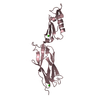
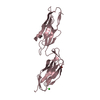
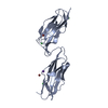
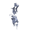
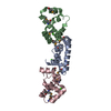
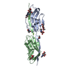
| Title | Crystal structure of a sHIP (UniProt Id: Q99XU0) mutant from Streptococcus pyogenes | ||||||
 Components Components | sHIP | ||||||
 Keywords Keywords | UNKNOWN FUNCTION /  helix-loop-helix helix-loop-helix | ||||||
| Function / homology | Protein of unknown function DUF4298 / Domain of unknown function (DUF4298) / identical protein binding / metal ion binding / DUF4298 domain-containing protein Function and homology information Function and homology information | ||||||
| Biological species |  Streptococcus pyogenes serotype M1 (bacteria) Streptococcus pyogenes serotype M1 (bacteria) | ||||||
| Method | X-RAY DIFFRACTION / SYNCHROTRON / MOLECULAR REPLACEMENT / Resolution: 1.73 Å | ||||||
 Authors Authors | Wisniewska, M. / Happonen, L. / Frick, I.-M. / Bjorck, L. / Streicher, W. / Malmstrom, J. / Wikstrom, M. | ||||||
 Citation Citation | Journal: J.Biol.Chem. / Year: 2014 Title: Functional and structural properties of a novel protein and virulence factor (Protein sHIP) in Streptococcus pyogenes. Authors: Wisniewska, M. / Happonen, L. / Kahn, F. / Varjosalo, M. / Malmstrom, L. / Rosenberger, G. / Karlsson, C. / Cazzamali, G. / Pozdnyakova, I. / Frick, I.M. / Bjorck, L. / Streicher, W. / ...Authors: Wisniewska, M. / Happonen, L. / Kahn, F. / Varjosalo, M. / Malmstrom, L. / Rosenberger, G. / Karlsson, C. / Cazzamali, G. / Pozdnyakova, I. / Frick, I.M. / Bjorck, L. / Streicher, W. / Malmstrom, J. / Wikstrom, M. | ||||||
| History |
|
- Structure visualization
| Structure viewer | Molecule: MolmilJmol/JSmol |
|---|
- Downloads & links
-Download
| PDBx/mmCIF format | 4pz1.cif.gz | 34.5 KB | Display | PDBx/mmCIF format |
|---|---|---|---|---|
| PDB format | pdb4pz1.ent.gz | 22.7 KB | Display | PDB format |
| PDBx/mmJSON format | 4pz1.json.gz | Tree view | PDBx/mmJSON format | |
| Others |  Other downloads Other downloads |
-Validation report
| Arichive directory | https://data.pdbj.org/pub/pdb/validation_reports/pz/4pz1ftp://data.pdbj.org/pub/pdb/validation_reports/pz/4pz1 | HTTPS FTP |
|---|
-Related structure data
| Related structure data |  4merSC S: Starting model for refinement C: citing same article ( |
|---|---|
| Similar structure data |


-Links
 PDBj
PDBj- Assembly
| Deposited unit | 
| ||||||||
|---|---|---|---|---|---|---|---|---|---|
| 1 | 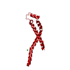
| ||||||||
| Unit cell |
|
-Components
| #1: Protein | Mass: 11350.693 Da / Num. of mol.: 1 / Mutation: C65S, L84A, L88A, Y95A Source method: isolated from a genetically manipulated source Source: (gene. exp.) Streptococcus pyogenes serotype M1 (bacteria)Gene: M5005_Spy1730, SPy_2034 / Production host: Escherichia coli (E. coli) / References: UniProt: Q99XU0 | ||||
|---|---|---|---|---|---|
| #2: Chemical | Chloride  Mass: 35.453 Da / Num. of mol.: 2 / Source method: obtained synthetically / Formula: Cl Mass: 35.453 Da / Num. of mol.: 2 / Source method: obtained synthetically / Formula: Cl#3: Chemical | ChemComp-CA / |   Mass: 40.078 Da / Num. of mol.: 1 / Source method: obtained synthetically / Formula: Ca Mass: 40.078 Da / Num. of mol.: 1 / Source method: obtained synthetically / Formula: Ca#4: Water | ChemComp-HOH / | Water Mass: 18.015 Da / Num. of mol.: 78 / Source method: isolated from a natural source / Formula: H2O Mass: 18.015 Da / Num. of mol.: 78 / Source method: isolated from a natural source / Formula: H2O |
-Experimental details
-Experiment
| Experiment | Method: X-RAY DIFFRACTION / Number of used crystals: 1 |
|---|
- Sample preparation
| Crystal | Density Matthews: 1.93 Å3/Da / Density % sol: 36.22 % |
|---|---|
| Crystal grow | Temperature: 291 K / Method: vapor diffusion, sitting drop / pH: 8 Details: 20% PEG 6000, 0.2 M calcium chloride, 0.1 M Tris, pH 8.0, VAPOR DIFFUSION, SITTING DROP, temperature 291K |
-Data collection
| Diffraction | Mean temperature: 100 K |
|---|---|
| Diffraction source | Source: SYNCHROTRON / Site: MAX II  / Beamline: I911-2 / Wavelength: 1.03841 Å / Beamline: I911-2 / Wavelength: 1.03841 Å |
| Detector | Type: MAR CCD 165 mm / Detector: CCD / Date: Dec 12, 2013 |
| Radiation | Monochromator: Bent Si (111) crystal, horizontally focusing / Protocol: SINGLE WAVELENGTH / Monochromatic (M) / Laue (L): M / Scattering type: x-ray |
| Radiation wavelength | Wavelength: 1.03841 Å / Relative weight: 1 |
| Reflection | Resolution: 1.73→19.76 Å / Num. obs: 9571 / % possible obs: 98.8 % / Rmerge(I) obs: 0.064 |
| Reflection shell | Resolution: 1.73→1.82 Å / % possible all: 97.8 |
- Processing
| Software |
| ||||||||||||||||||||||||||||||||||||||||||||||||||||||||||||
|---|---|---|---|---|---|---|---|---|---|---|---|---|---|---|---|---|---|---|---|---|---|---|---|---|---|---|---|---|---|---|---|---|---|---|---|---|---|---|---|---|---|---|---|---|---|---|---|---|---|---|---|---|---|---|---|---|---|---|---|---|---|
| Refinement | Method to determine structure: MOLECULAR REPLACEMENT Starting model: PDB entry 4MER Resolution: 1.73→19.76 Å / Cor.coef. Fo:Fc: 0.945 / Cor.coef. Fo:Fc free: 0.935 / SU B: 2.071 / SU ML: 0.069 / Cross valid method: THROUGHOUT / ESU R: 0.124 / ESU R Free: 0.116 / Stereochemistry target values: MAXIMUM LIKELIHOOD / Details: HYDROGENS HAVE BEEN ADDED IN THE RIDING POSITIONS
| ||||||||||||||||||||||||||||||||||||||||||||||||||||||||||||
| Solvent computation | Ion probe radii: 0.8 Å / Shrinkage radii: 0.8 Å / VDW probe radii: 1.2 Å / Solvent model: MASK | ||||||||||||||||||||||||||||||||||||||||||||||||||||||||||||
| Displacement parameters | Biso mean: 12.789 Å2
| ||||||||||||||||||||||||||||||||||||||||||||||||||||||||||||
| Refinement step | Cycle: LAST / Resolution: 1.73→19.76 Å
| ||||||||||||||||||||||||||||||||||||||||||||||||||||||||||||
| Refine LS restraints |
| ||||||||||||||||||||||||||||||||||||||||||||||||||||||||||||
| LS refinement shell | Resolution: 1.73→1.775 Å / Total num. of bins used: 20
|
