 Movie
Movie Controller


+ Open data

- Basic information
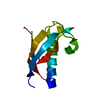
| Entry | Database: PDB / ID: 7cn6 | |||||||||
|---|---|---|---|---|---|---|---|---|---|---|
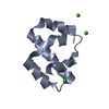
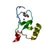
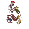
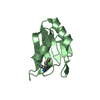
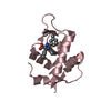
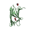
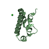
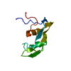
| Title | T4 phage spackle protein gp61.3 | |||||||||
 Components Components | Protein spackle | |||||||||
 Keywords Keywords |  VIRAL PROTEIN / Lysozyme inhibitor / Phage Lysis inhibition VIRAL PROTEIN / Lysozyme inhibitor / Phage Lysis inhibition | |||||||||
| Function / homology | Protein spackle / superinfection exclusion / host cell periplasmic space / Protein spackle Function and homology information Function and homology information | |||||||||
| Biological species |  Enterobacteria phage T4 (virus) Enterobacteria phage T4 (virus) | |||||||||
| Method | X-RAY DIFFRACTION / SYNCHROTRON / SAD / Resolution: 1.6 Å | |||||||||
 Authors Authors | Kanamaru, S. / Leiman, P.G. | |||||||||
| Funding support |  Japan, 2items Japan, 2items
| |||||||||
 Citation Citation | Journal: Viruses / Year: 2020 Title: Structure and Function of the T4 Spackle Protein Gp61.3. Authors: Kanamaru, S. / Uchida, K. / Nemoto, M. / Fraser, A. / Arisaka, F. / Leiman, P.G. | |||||||||
| History |
|
- Structure visualization
| Structure viewer | Molecule: MolmilJmol/JSmol |
|---|
- Downloads & links
-Download
| PDBx/mmCIF format | 7cn6.cif.gz | 138.3 KB | Display | PDBx/mmCIF format |
|---|---|---|---|---|
| PDB format | pdb7cn6.ent.gz | 95.8 KB | Display | PDB format |
| PDBx/mmJSON format | 7cn6.json.gz | Tree view | PDBx/mmJSON format | |
| Others |  Other downloads Other downloads |
-Validation report
| Arichive directory | https://data.pdbj.org/pub/pdb/validation_reports/cn/7cn6ftp://data.pdbj.org/pub/pdb/validation_reports/cn/7cn6 | HTTPS FTP |
|---|
-Related structure data


-Links
 PDBj
PDBj- Assembly
| Deposited unit | 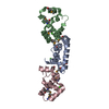
| ||||||||||||
|---|---|---|---|---|---|---|---|---|---|---|---|---|---|
| 1 | 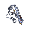
| ||||||||||||
| 2 | 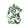
| ||||||||||||
| 3 | 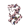
| ||||||||||||
| Unit cell |
|
-Components
| #1: Protein | Mass: 8876.365 Da / Num. of mol.: 3 Source method: isolated from a genetically manipulated source Details: SF file contains Friedel pairs. / Source: (gene. exp.) Enterobacteria phage T4 (virus) / Gene: sp, 61.3 / Plasmid: pUC19 / Production host:  Escherichia coli BL21(DE3) (bacteria) / References: UniProt: P39230 Escherichia coli BL21(DE3) (bacteria) / References: UniProt: P39230#2: Chemical |   Mass: 40.078 Da / Num. of mol.: 3 / Source method: obtained synthetically / Formula: Ca Mass: 40.078 Da / Num. of mol.: 3 / Source method: obtained synthetically / Formula: Ca#3: Water | ChemComp-HOH / | Water Mass: 18.015 Da / Num. of mol.: 399 / Source method: isolated from a natural source / Formula: H2O Mass: 18.015 Da / Num. of mol.: 399 / Source method: isolated from a natural source / Formula: H2OHas ligand of interest | N | |
|---|
-Experimental details
-Experiment
| Experiment | Method: X-RAY DIFFRACTION / Number of used crystals: 1 |
|---|
- Sample preparation
| Crystal | Density Matthews: 2.01 Å3/Da / Density % sol: 38.86 % |
|---|---|
| Crystal grow | Temperature: 293.15 K / Method: vapor diffusion, hanging drop / pH: 9.4 / Details: PEG4000, glycine, calcium chloride |
-Data collection
| Diffraction | Mean temperature: 100 K / Serial crystal experiment: N |
|---|---|
| Diffraction source | Source: SYNCHROTRON / Site: ESRF  / Beamline: ID30B / Wavelength: 0.9794 Å / Beamline: ID30B / Wavelength: 0.9794 Å |
| Detector | Type: ADSC QUANTUM 315 / Detector: CCD / Date: Sep 18, 2009 |
| Radiation | Protocol: MAD / Monochromatic (M) / Laue (L): M / Scattering type: x-ray |
| Radiation wavelength | Wavelength: 0.9794 Å / Relative weight: 1 |
| Reflection | Resolution: 1.6→39.8 Å / Num. obs: 51366 / % possible obs: 95.2 % / Redundancy: 1.9 % / Biso Wilson estimate: 11.76 Å2 / CC1/2: 0.979 / Rmerge(I) obs: 0.107 / Rpim(I) all: 0.107 / Rrim(I) all: 0.151 / Χ2: 0.83 / Net I/σ(I): 4 |
| Reflection shell | Resolution: 1.6→1.63 Å / Rmerge(I) obs: 0.19 / Mean I/σ(I) obs: 2.1 / Num. unique obs: 1001 / CC1/2: 0.895 / Rpim(I) all: 0.19 / Rrim(I) all: 0.269 / Χ2: 0.47 / % possible all: 75.4 |
- Processing
| Software |
| |||||||||||||||||||||||||||||||||||||||||||||||||||||||||||||||||||||||||||||||||||||||||||||||||||||||||||||||||||||||||||||||||||||
|---|---|---|---|---|---|---|---|---|---|---|---|---|---|---|---|---|---|---|---|---|---|---|---|---|---|---|---|---|---|---|---|---|---|---|---|---|---|---|---|---|---|---|---|---|---|---|---|---|---|---|---|---|---|---|---|---|---|---|---|---|---|---|---|---|---|---|---|---|---|---|---|---|---|---|---|---|---|---|---|---|---|---|---|---|---|---|---|---|---|---|---|---|---|---|---|---|---|---|---|---|---|---|---|---|---|---|---|---|---|---|---|---|---|---|---|---|---|---|---|---|---|---|---|---|---|---|---|---|---|---|---|---|---|---|
| Refinement | Method to determine structure: SAD / Resolution: 1.6→19.9 Å / SU ML: 0.1797 / Cross valid method: FREE R-VALUE / σ(F): 1.94 / Phase error: 28.4219 Stereochemistry target values: GeoStd + Monomer Library + CDL v1.2
| |||||||||||||||||||||||||||||||||||||||||||||||||||||||||||||||||||||||||||||||||||||||||||||||||||||||||||||||||||||||||||||||||||||
| Solvent computation | Shrinkage radii: 0.9 Å / VDW probe radii: 1.11 Å / Solvent model: FLAT BULK SOLVENT MODEL | |||||||||||||||||||||||||||||||||||||||||||||||||||||||||||||||||||||||||||||||||||||||||||||||||||||||||||||||||||||||||||||||||||||
| Displacement parameters | Biso mean: 17.96 Å2 | |||||||||||||||||||||||||||||||||||||||||||||||||||||||||||||||||||||||||||||||||||||||||||||||||||||||||||||||||||||||||||||||||||||
| Refinement step | Cycle: LAST / Resolution: 1.6→19.9 Å
| |||||||||||||||||||||||||||||||||||||||||||||||||||||||||||||||||||||||||||||||||||||||||||||||||||||||||||||||||||||||||||||||||||||
| Refine LS restraints |
| |||||||||||||||||||||||||||||||||||||||||||||||||||||||||||||||||||||||||||||||||||||||||||||||||||||||||||||||||||||||||||||||||||||
| LS refinement shell |
|
