 Movie
Movie Controller

[English] 日本語
 Yorodumi
- PDB-4obv: Ruminococcus gnavus tryptophan decarboxylase RUMGNA_01526 (alpha-FMT) -
+ Open data

- Basic information
| Entry | Database: PDB / ID: 4obv | ||||||
|---|---|---|---|---|---|---|---|
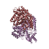
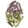
| Title | Ruminococcus gnavus tryptophan decarboxylase RUMGNA_01526 (alpha-FMT) | ||||||
 Components Components | Pyridoxal-dependent decarboxylase domain protein | ||||||
 Keywords Keywords |  LYASE / Type 1 PLP-dependent / Decarboxylase LYASE / Type 1 PLP-dependent / Decarboxylase | ||||||
| Function / homology |  Function and homology informationL-tryptophan decarboxylase / L-tryptophan decarboxylase activity / tryptophan metabolic process / pyridoxal phosphate binding / cytoplasm Function and homology informationL-tryptophan decarboxylase / L-tryptophan decarboxylase activity / tryptophan metabolic process / pyridoxal phosphate binding / cytoplasmSimilarity search - Function | ||||||
| Biological species |  Ruminococcus gnavus (bacteria) Ruminococcus gnavus (bacteria) | ||||||
| Method | X-RAY DIFFRACTION / SYNCHROTRON / MOLECULAR REPLACEMENT / Resolution: 2.84 Å | ||||||
 Authors Authors | Fraser, J.S. / Van Benschoten, A.H. | ||||||
 Citation Citation | Journal: Cell Host Microbe / Year: 2014 Title: Discovery and Characterization of Gut Microbiota Decarboxylases that Can Produce the Neurotransmitter Tryptamine. Authors: Williams, B.B. / Van Benschoten, A.H. / Cimermancic, P. / Donia, M.S. / Zimmermann, M. / Taketani, M. / Ishihara, A. / Kashyap, P.C. / Fraser, J.S. / Fischbach, M.A. | ||||||
| History |
|
- Structure visualization
| Structure viewer | Molecule: MolmilJmol/JSmol |
|---|
- Downloads & links
-Download
| PDBx/mmCIF format | 4obv.cif.gz | 370.8 KB | Display | PDBx/mmCIF format |
|---|---|---|---|---|
| PDB format | pdb4obv.ent.gz | 305.7 KB | Display | PDB format |
| PDBx/mmJSON format | 4obv.json.gz | Tree view | PDBx/mmJSON format | |
| Others |  Other downloads Other downloads |
-Validation report
| Arichive directory | https://data.pdbj.org/pub/pdb/validation_reports/ob/4obvftp://data.pdbj.org/pub/pdb/validation_reports/ob/4obv | HTTPS FTP |
|---|
-Related structure data


-Links
 PDBj
PDBj- Assembly
| Deposited unit | 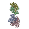
| |||||||||
|---|---|---|---|---|---|---|---|---|---|---|
| 1 | 
| |||||||||
| 2 | 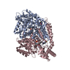
| |||||||||
| Unit cell |
| |||||||||
| Noncrystallographic symmetry (NCS) | NCS domain:
|
-Components
| #1: Protein | Mass: 55024.621 Da / Num. of mol.: 4 Source method: isolated from a genetically manipulated source Source: (gene. exp.) Ruminococcus gnavus (bacteria) / Gene: RUMGNA_01526 / Production host: Escherichia coli (E. coli) / References: UniProt: A7B1V0#2: Chemical | ChemComp-2SU /   Mass: 236.242 Da / Num. of mol.: 4 / Source method: obtained synthetically / Formula: C12H13FN2O2 Mass: 236.242 Da / Num. of mol.: 4 / Source method: obtained synthetically / Formula: C12H13FN2O2#3: Chemical | ChemComp-3SO / {   Mass: 402.338 Da / Num. of mol.: 4 / Source method: obtained synthetically / Formula: C19H19N2O6P Mass: 402.338 Da / Num. of mol.: 4 / Source method: obtained synthetically / Formula: C19H19N2O6P |
|---|
-Experimental details
-Experiment
| Experiment | Method: X-RAY DIFFRACTION / Number of used crystals: 1 |
|---|
- Sample preparation
| Crystal | Density Matthews: 2.59 Å3/Da / Density % sol: 52.45 % |
|---|---|
| Crystal grow | Temperature: 298 K / Method: vapor diffusion, hanging drop / pH: 5.25 Details: 30% ethoxyethanol, 0.1M citrate pH 5.25, 4% polypropylene P400, VAPOR DIFFUSION, HANGING DROP, temperature 298K |
-Data collection
| Diffraction | Mean temperature: 100 K |
|---|---|
| Diffraction source | Source: SYNCHROTRON / Site: ALS  / Beamline: 8.3.1 / Wavelength: 1.1159 Å / Beamline: 8.3.1 / Wavelength: 1.1159 Å |
| Detector | Type: ADSC QUANTUM 315r / Detector: CCD / Date: Jan 9, 2013 |
| Radiation | Monochromator: Double flat crystal, Si 111 / Protocol: SINGLE WAVELENGTH / Monochromatic (M) / Laue (L): M / Scattering type: x-ray |
| Radiation wavelength | Wavelength: 1.1159 Å / Relative weight: 1 |
| Reflection | Resolution: 2.84→52.442 Å / Num. all: 55262 / Num. obs: 55229 / % possible obs: 99.94 % / Observed criterion σ(F): 0 / Observed criterion σ(I): 0 |
| Reflection shell | Resolution: 2.84→2.991 Å / % possible all: 99.94 |
- Processing
| Software |
| |||||||||||||||||||||||||||||||||||||||||||||||||||||||||||||||||||||||||||||||||||||||||||||||||||||||||
|---|---|---|---|---|---|---|---|---|---|---|---|---|---|---|---|---|---|---|---|---|---|---|---|---|---|---|---|---|---|---|---|---|---|---|---|---|---|---|---|---|---|---|---|---|---|---|---|---|---|---|---|---|---|---|---|---|---|---|---|---|---|---|---|---|---|---|---|---|---|---|---|---|---|---|---|---|---|---|---|---|---|---|---|---|---|---|---|---|---|---|---|---|---|---|---|---|---|---|---|---|---|---|---|---|---|---|
| Refinement | Method to determine structure: MOLECULAR REPLACEMENT / Resolution: 2.84→52.442 Å / SU ML: 0.39 / σ(F): 1.34 / Phase error: 24.72 / Stereochemistry target values: ML
| |||||||||||||||||||||||||||||||||||||||||||||||||||||||||||||||||||||||||||||||||||||||||||||||||||||||||
| Solvent computation | Shrinkage radii: 0.9 Å / VDW probe radii: 1.11 Å / Solvent model: FLAT BULK SOLVENT MODEL | |||||||||||||||||||||||||||||||||||||||||||||||||||||||||||||||||||||||||||||||||||||||||||||||||||||||||
| Refinement step | Cycle: LAST / Resolution: 2.84→52.442 Å
| |||||||||||||||||||||||||||||||||||||||||||||||||||||||||||||||||||||||||||||||||||||||||||||||||||||||||
| Refine LS restraints |
| |||||||||||||||||||||||||||||||||||||||||||||||||||||||||||||||||||||||||||||||||||||||||||||||||||||||||
| LS refinement shell |
|
