 Movie
Movie Controller


+ Open data

- Basic information
| Entry | Database: PDB / ID: 6liv | |||||||||
|---|---|---|---|---|---|---|---|---|---|---|
| Title | Crystal structure of Tyrosine decarboxylase in complex with PLP | |||||||||
 Components Components | Tyrosine/DOPA decarboxylase 2 | |||||||||
 Keywords Keywords |  LYASE / holo form / decarboxylase / PLP binding / tyrosine LYASE / holo form / decarboxylase / PLP binding / tyrosine | |||||||||
| Function / homology |  Function and homology information Function and homology informationL-dopa decarboxylase activity / 5-hydroxy-L-tryptophan decarboxylase activity / aromatic-L-amino-acid decarboxylase / tyrosine decarboxylase / tyrosine decarboxylase activity / amino acid metabolic process / pyridoxal phosphate bindingSimilarity search - Function | |||||||||
| Biological species |  Papaver somniferum (opium poppy) Papaver somniferum (opium poppy) | |||||||||
| Method | X-RAY DIFFRACTION / SYNCHROTRON / MOLECULAR REPLACEMENT / Resolution: 2.31 Å | |||||||||
 Authors Authors | Wang, H. / Yu, J. / Yao, M. | |||||||||
| Funding support |  Japan, 2items Japan, 2items
| |||||||||
 Citation Citation | Journal: Biochem.Biophys.Res.Commun. / Year: 2019 Title: Crystal structures clarify cofactor binding of plant tyrosine decarboxylase. Authors: Wang, H. / Yu, J. / Satoh, Y. / Nakagawa, Y. / Tanaka, R. / Kato, K. / Yao, M. | |||||||||
| History |
|
- Structure visualization
| Structure viewer | Molecule: MolmilJmol/JSmol |
|---|
- Downloads & links
-Download
| PDBx/mmCIF format | 6liv.cif.gz | 635 KB | Display | PDBx/mmCIF format |
|---|---|---|---|---|
| PDB format | pdb6liv.ent.gz | 479.6 KB | Display | PDB format |
| PDBx/mmJSON format | 6liv.json.gz | Tree view | PDBx/mmJSON format | |
| Others |  Other downloads Other downloads |
-Validation report
| Arichive directory | https://data.pdbj.org/pub/pdb/validation_reports/li/6livftp://data.pdbj.org/pub/pdb/validation_reports/li/6liv | HTTPS FTP |
|---|
-Related structure data
| Related structure data |  6liuC  1js3S S: Starting model for refinement C: citing same article ( |
|---|---|
| Similar structure data |


-Links
 PDBj
PDBj- Assembly
| Deposited unit | 
| ||||||||||||
|---|---|---|---|---|---|---|---|---|---|---|---|---|---|
| 1 | 
| ||||||||||||
| 2 | 
| ||||||||||||
| 3 | 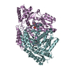
| ||||||||||||
| Unit cell |
|
-Components
| #1: Protein | Mass: 59651.719 Da / Num. of mol.: 6 Source method: isolated from a genetically manipulated source Source: (gene. exp.) Papaver somniferum (opium poppy) / Gene: TYDC2 / Production host:  Escherichia coli (E. coli) Escherichia coli (E. coli)References: UniProt: P54769, aromatic-L-amino-acid decarboxylase, tyrosine decarboxylase#2: Chemical | ChemComp-GOL / Glycerol  Mass: 92.094 Da / Num. of mol.: 9 / Source method: isolated from a natural source / Formula: C3H8O3 Mass: 92.094 Da / Num. of mol.: 9 / Source method: isolated from a natural source / Formula: C3H8O3#3: Water | ChemComp-HOH / | Water Mass: 18.015 Da / Num. of mol.: 1018 / Source method: isolated from a natural source / Formula: H2O Mass: 18.015 Da / Num. of mol.: 1018 / Source method: isolated from a natural source / Formula: H2OHas ligand of interest | Y | |
|---|
-Experimental details
-Experiment
| Experiment | Method: X-RAY DIFFRACTION / Number of used crystals: 1 |
|---|
- Sample preparation
| Crystal | Density Matthews: 3.24 Å3/Da / Density % sol: 62.04 % |
|---|---|
| Crystal grow | Temperature: 293 K / Method: vapor diffusion, hanging drop / Details: disodium malonate, HEPES, TCEP-HCl, glycerol |
-Data collection
| Diffraction | Mean temperature: 80 K / Serial crystal experiment: N |
|---|---|
| Diffraction source | Source: SYNCHROTRON / Site: Photon Factory / Beamline: BL-1A / Wavelength: 1.1 Å |
| Detector | Type: DECTRIS EIGER X 16M / Detector: PIXEL / Date: Dec 15, 2018 |
| Radiation | Protocol: SINGLE WAVELENGTH / Monochromatic (M) / Laue (L): M / Scattering type: x-ray |
| Radiation wavelength | Wavelength: 1.1 Å / Relative weight: 1 |
| Reflection | Resolution: 2.31→50 Å / % possible obs: 99.7 % / Redundancy: 6.96 % / Biso Wilson estimate: 32.07 Å2 / CC1/2: 0.991 / Rrim(I) all: 0.214 / Net I/σ(I): 7.12 |
| Reflection shell | Resolution: 2.31→2.45 Å / Redundancy: 6.66 % / Mean I/σ(I) obs: 1.7 / Num. unique obs: 31999 / CC1/2: 0.712 / Rrim(I) all: 0.887 / % possible all: 98.4 |
- Processing
| Software |
| |||||||||||||||||||||||||||||||||||||||||||||||||||||||||||||||||||||||||||||||||||||||||||||||||||||||||||||||||||||||||||||||||||||||||||||||||||||||||||||||||||||||||||||||||||||||||||||||||||||||||||||||||||||||||
|---|---|---|---|---|---|---|---|---|---|---|---|---|---|---|---|---|---|---|---|---|---|---|---|---|---|---|---|---|---|---|---|---|---|---|---|---|---|---|---|---|---|---|---|---|---|---|---|---|---|---|---|---|---|---|---|---|---|---|---|---|---|---|---|---|---|---|---|---|---|---|---|---|---|---|---|---|---|---|---|---|---|---|---|---|---|---|---|---|---|---|---|---|---|---|---|---|---|---|---|---|---|---|---|---|---|---|---|---|---|---|---|---|---|---|---|---|---|---|---|---|---|---|---|---|---|---|---|---|---|---|---|---|---|---|---|---|---|---|---|---|---|---|---|---|---|---|---|---|---|---|---|---|---|---|---|---|---|---|---|---|---|---|---|---|---|---|---|---|---|---|---|---|---|---|---|---|---|---|---|---|---|---|---|---|---|---|---|---|---|---|---|---|---|---|---|---|---|---|---|---|---|---|---|---|---|---|---|---|---|---|---|---|---|---|---|---|---|---|
| Refinement | Method to determine structure: MOLECULAR REPLACEMENT Starting model: 1JS3 Resolution: 2.31→49.84 Å / SU ML: 0.28 / Cross valid method: FREE R-VALUE / σ(F): 1.34 / Phase error: 25.1863 Stereochemistry target values: GeoStd + Monomer Library + CDL v1.2
| |||||||||||||||||||||||||||||||||||||||||||||||||||||||||||||||||||||||||||||||||||||||||||||||||||||||||||||||||||||||||||||||||||||||||||||||||||||||||||||||||||||||||||||||||||||||||||||||||||||||||||||||||||||||||
| Solvent computation | Shrinkage radii: 0.9 Å / VDW probe radii: 1.11 Å / Solvent model: FLAT BULK SOLVENT MODEL | |||||||||||||||||||||||||||||||||||||||||||||||||||||||||||||||||||||||||||||||||||||||||||||||||||||||||||||||||||||||||||||||||||||||||||||||||||||||||||||||||||||||||||||||||||||||||||||||||||||||||||||||||||||||||
| Displacement parameters | Biso mean: 45.32 Å2 | |||||||||||||||||||||||||||||||||||||||||||||||||||||||||||||||||||||||||||||||||||||||||||||||||||||||||||||||||||||||||||||||||||||||||||||||||||||||||||||||||||||||||||||||||||||||||||||||||||||||||||||||||||||||||
| Refinement step | Cycle: LAST / Resolution: 2.31→49.84 Å
| |||||||||||||||||||||||||||||||||||||||||||||||||||||||||||||||||||||||||||||||||||||||||||||||||||||||||||||||||||||||||||||||||||||||||||||||||||||||||||||||||||||||||||||||||||||||||||||||||||||||||||||||||||||||||
| Refine LS restraints |
| |||||||||||||||||||||||||||||||||||||||||||||||||||||||||||||||||||||||||||||||||||||||||||||||||||||||||||||||||||||||||||||||||||||||||||||||||||||||||||||||||||||||||||||||||||||||||||||||||||||||||||||||||||||||||
| LS refinement shell |
|
