 Movie
Movie Controller


+ Open data

- Basic information
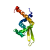
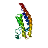
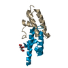
| Entry | Database: PDB / ID: 4n6l | ||||||
|---|---|---|---|---|---|---|---|
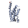
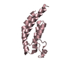
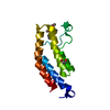
| Title | Crystal structure of human cystatin E/M | ||||||
 Components Components | Cystatin-M | ||||||
 Keywords Keywords | HYDROLASE INHIBITOR /  cysteine protease inhibitor / legumain / asparaginyl endopeptidase / reactive center loop / papain / cathepsin / cancer / cystatin fold / protease inhibitor cysteine protease inhibitor / legumain / asparaginyl endopeptidase / reactive center loop / papain / cathepsin / cancer / cystatin fold / protease inhibitor | ||||||
| Function / homology |  Function and homology information Function and homology informationcornified envelope / cysteine-type endopeptidase inhibitor activity / anatomical structure morphogenesis / epidermis development / extracellular exosome Similarity search - Function | ||||||
| Biological species |  Homo sapiens (human) Homo sapiens (human) | ||||||
| Method | X-RAY DIFFRACTION / SYNCHROTRON / MOLECULAR REPLACEMENT / Resolution: 1.952 Å | ||||||
 Authors Authors | Dall, E. / Brandstetter, H. | ||||||
 Citation Citation | Journal: Angew.Chem.Int.Ed.Engl. / Year: 2015 Title: Structure and mechanism of an aspartimide-dependent Peptide ligase in human legumain. Authors: Dall, E. / Fegg, J.C. / Briza, P. / Brandstetter, H. | ||||||
| History |
|
- Structure visualization
| Structure viewer | Molecule: MolmilJmol/JSmol |
|---|
- Downloads & links
-Download
| PDBx/mmCIF format | 4n6l.cif.gz | 36.9 KB | Display | PDBx/mmCIF format |
|---|---|---|---|---|
| PDB format | pdb4n6l.ent.gz | 24.4 KB | Display | PDB format |
| PDBx/mmJSON format | 4n6l.json.gz | Tree view | PDBx/mmJSON format | |
| Others |  Other downloads Other downloads |
-Validation report
| Arichive directory | https://data.pdbj.org/pub/pdb/validation_reports/n6/4n6lftp://data.pdbj.org/pub/pdb/validation_reports/n6/4n6l | HTTPS FTP |
|---|
-Related structure data
| Related structure data | 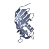 4n6mC  4n6nC  4n6oC  3gaxS C: citing same article ( S: Starting model for refinement |
|---|---|
| Similar structure data |


-Links
 PDBj
PDBj

- Assembly
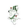
| Deposited unit | 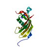
| ||||||||
|---|---|---|---|---|---|---|---|---|---|
| 1 |
| ||||||||
| Unit cell |
|
-Components
| #1: Protein | Mass: 14924.931 Da / Num. of mol.: 1 / Fragment: UNP residues 29-149 Source method: isolated from a genetically manipulated source Source: (gene. exp.) Homo sapiens (human) / Gene: CST6 / Production host:  Escherichia coli (E. coli) / References: UniProt: Q15828 Escherichia coli (E. coli) / References: UniProt: Q15828 |
|---|---|
| #2: Water | ChemComp-HOH / Water Mass: 18.015 Da / Num. of mol.: 91 / Source method: isolated from a natural source / Formula: H2O Mass: 18.015 Da / Num. of mol.: 91 / Source method: isolated from a natural source / Formula: H2O |
-Experimental details
-Experiment
| Experiment | Method: X-RAY DIFFRACTION / Number of used crystals: 1 |
|---|
- Sample preparation
| Crystal | Density Matthews: 1.98 Å3/Da / Density % sol: 37.81 % |
|---|---|
| Crystal grow | Temperature: 293 K / Method: vapor diffusion, sitting drop / pH: 4.6 Details: 30 % PEG 4000, 100 mM sodium acetate pH 4.6, 200 mM ammonium sulfate, VAPOR DIFFUSION, SITTING DROP, temperature 293K |
-Data collection
| Diffraction | Mean temperature: 100 K |
|---|---|
| Diffraction source | Source: SYNCHROTRON / Site: ESRF  / Beamline: ID29 / Wavelength: 0.9763 Å / Beamline: ID29 / Wavelength: 0.9763 Å |
| Detector | Type: DECTRIS PILATUS 6M / Detector: PIXEL / Date: Jul 24, 2012 |
| Radiation | Monochromator: Si(111) / Protocol: SINGLE WAVELENGTH / Monochromatic (M) / Laue (L): M / Scattering type: x-ray |
| Radiation wavelength | Wavelength: 0.9763 Å / Relative weight: 1 |
| Reflection | Resolution: 1.95→68.6 Å / Num. obs: 8955 / % possible obs: 98.9 % / Observed criterion σ(F): 2 / Observed criterion σ(I): 2 |
| Reflection shell | Resolution: 1.95→2.06 Å / % possible all: 98.8 |
- Processing
| Software |
| ||||||||||||||||||||||||||||
|---|---|---|---|---|---|---|---|---|---|---|---|---|---|---|---|---|---|---|---|---|---|---|---|---|---|---|---|---|---|
| Refinement | Method to determine structure: MOLECULAR REPLACEMENT Starting model: PDB ENTRY 3GAX Resolution: 1.952→36.269 Å / SU ML: 0.17 / σ(F): 1.38 / Phase error: 27.59 / Stereochemistry target values: ML
| ||||||||||||||||||||||||||||
| Solvent computation | Shrinkage radii: 0.9 Å / VDW probe radii: 1.11 Å / Solvent model: FLAT BULK SOLVENT MODEL | ||||||||||||||||||||||||||||
| Refinement step | Cycle: LAST / Resolution: 1.952→36.269 Å
| ||||||||||||||||||||||||||||
| Refine LS restraints |
| ||||||||||||||||||||||||||||
| LS refinement shell |
|
