 Movie
Movie Controller

[English] 日本語
 Yorodumi
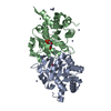
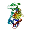
- PDB-4n6o: Crystal structure of reduced legumain in complex with cystatin E/M -
+ Open data

- Basic information
| Entry | Database: PDB / ID: 4n6o | ||||||||||||
|---|---|---|---|---|---|---|---|---|---|---|---|---|---|
| Title | Crystal structure of reduced legumain in complex with cystatin E/M | ||||||||||||
 Components Components |
| ||||||||||||
 Keywords Keywords | HYDROLASE/HYDROLASE INHIBITOR /  complex / cysteine protease / inhibitor / legumain / asparaginyl endopeptidase / reactive center loop / papain / cathepsin / cancer / HYDROLASE-HYDROLASE INHIBITOR complex complex / cysteine protease / inhibitor / legumain / asparaginyl endopeptidase / reactive center loop / papain / cathepsin / cancer / HYDROLASE-HYDROLASE INHIBITOR complex | ||||||||||||
| Function / homology |  Function and homology information Function and homology informationnegative regulation of ERBB signaling pathway / legumain / vacuolar protein processing / renal system process / Vitamin D (calciferol) metabolism / receptor catabolic process / vitamin D metabolic process / self proteolysis / cornified envelope / endolysosome lumen ...negative regulation of ERBB signaling pathway / legumain / vacuolar protein processing / renal system process / Vitamin D (calciferol) metabolism / receptor catabolic process / vitamin D metabolic process / self proteolysis / cornified envelope / endolysosome lumen / activation of cysteine-type endopeptidase activity / response to acidic pH / dendritic spine organization / positive regulation of monocyte chemotaxis / Trafficking and processing of endosomal TLR / positive regulation of endothelial cell chemotaxis / negative regulation of multicellular organism growth / cysteine-type endopeptidase inhibitor activity / cellular response to hepatocyte growth factor stimulus / associative learning / protein maturation / endopeptidase activator activity / anatomical structure morphogenesis / epidermis development / cellular response to calcium ion / MHC class II antigen presentation / positive regulation of mitotic cell cycle / lysosomal lumen / proteolysis involved in protein catabolic process / positive regulation of long-term synaptic potentiation / tau protein binding / memory / cellular response to amyloid-beta / antigen processing and presentation of exogenous peptide antigen via MHC class II / late endosome / apical part of cell / peptidase activity / negative regulation of neuron apoptotic process / lysosome / cysteine-type endopeptidase activity / negative regulation of gene expression / positive regulation of cell population proliferation / perinuclear region of cytoplasm / proteolysis / extracellular exosome / extracellular region / cytoplasmSimilarity search - Function | ||||||||||||
| Biological species |  Homo sapiens (human) Homo sapiens (human) | ||||||||||||
| Method | X-RAY DIFFRACTION / SYNCHROTRON / MOLECULAR REPLACEMENT / Resolution: 1.8 Å | ||||||||||||
 Authors Authors | Dall, E. / Brandstetter, H. | ||||||||||||
 Citation Citation | Journal: Angew.Chem.Int.Ed.Engl. / Year: 2015 Title: Structure and mechanism of an aspartimide-dependent Peptide ligase in human legumain. Authors: Dall, E. / Fegg, J.C. / Briza, P. / Brandstetter, H. | ||||||||||||
| History |
| ||||||||||||
| Remark 650 | HELIX DETERMINATION METHOD: AUTHOR DETERMINED |
- Structure visualization
| Structure viewer | Molecule: MolmilJmol/JSmol |
|---|
- Downloads & links
-Download
| PDBx/mmCIF format | 4n6o.cif.gz | 98.9 KB | Display | PDBx/mmCIF format |
|---|---|---|---|---|
| PDB format | pdb4n6o.ent.gz | 72.7 KB | Display | PDB format |
| PDBx/mmJSON format | 4n6o.json.gz | Tree view | PDBx/mmJSON format | |
| Others |  Other downloads Other downloads |
-Validation report
| Arichive directory | https://data.pdbj.org/pub/pdb/validation_reports/n6/4n6oftp://data.pdbj.org/pub/pdb/validation_reports/n6/4n6o | HTTPS FTP |
|---|
-Related structure data
| Related structure data | 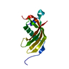 4n6lSC 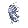 4n6mC  4n6nC 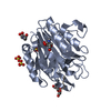 4aw9S S: Starting model for refinement C: citing same article ( |
|---|---|
| Similar structure data |


-Links
 PDBj
PDBj


- Assembly
| Deposited unit | 
| ||||||||
|---|---|---|---|---|---|---|---|---|---|
| 1 |
| ||||||||
| Unit cell |
|
-Components
-Protein , 2 types, 2 molecules AB
| #1: Protein | Asparagine endopeptidase / Asparaginyl endopeptidase / Protease / cysteine 1 Mass: 31643.551 Da / Num. of mol.: 1 / Fragment: UNP residues 26-303 / Mutation: N263Q Source method: isolated from a genetically manipulated source Source: (gene. exp.) Homo sapiens (human) / Gene: LGMN, PRSC1 / Production host:  Leishmania tarentolae (eukaryote) / References: UniProt: Q99538, legumain Leishmania tarentolae (eukaryote) / References: UniProt: Q99538, legumain |
|---|---|
| #2: Protein | Mass: 14924.931 Da / Num. of mol.: 1 / Fragment: UNP residues 29-149 Source method: isolated from a genetically manipulated source Source: (gene. exp.) Homo sapiens (human) / Gene: CST6 / Production host:  Escherichia coli (E. coli) / References: UniProt: Q15828 Escherichia coli (E. coli) / References: UniProt: Q15828 |
-Sugars , 2 types, 2 molecules
| #3: Polysaccharide | beta-D-mannopyranose-(1-4)-2-acetamido-2-deoxy-beta-D-glucopyranose-(1-4)-2-acetamido-2-deoxy-beta- ...beta-D-mannopyranose-(1-4)-2-acetamido-2-deoxy-beta-D-glucopyranose-(1-4)-2-acetamido-2-deoxy-beta-D-glucopyranose / Mass: 586.542 Da / Num. of mol.: 1 Source method: isolated from a genetically manipulated source |
|---|---|
| #4: Polysaccharide | 2-acetamido-2-deoxy-beta-D-glucopyranose-(1-4)-2-acetamido-2-deoxy-beta-D-glucopyranose / Mass: 424.401 Da / Num. of mol.: 1 Source method: isolated from a genetically manipulated source |
-Non-polymers , 2 types, 284 molecules 

| #5: Chemical | ChemComp-IOD / Iodide Mass: 126.904 Da / Num. of mol.: 5 / Source method: obtained synthetically / Formula: I Mass: 126.904 Da / Num. of mol.: 5 / Source method: obtained synthetically / Formula: I#6: Water | ChemComp-HOH / | WaterMass: 18.015 Da / Num. of mol.: 279 / Source method: isolated from a natural source / Formula: H2O |
|---|
-Details
| Sequence details | RESIDUES 147 AND 148 STARTED OUT AS ASP AND HIS. THE SIDECHAIN OF ASP ATTACKS THE FOLLOWING AMIDE ...RESIDUES 147 AND 148 STARTED OUT AS ASP AND HIS. THE SIDECHAIN OF ASP ATTACKS THE FOLLOWING AMIDE OF HIS TO FORM A SUCCINIMID |
|---|
-Experimental details
-Experiment
| Experiment | Method: X-RAY DIFFRACTION / Number of used crystals: 1 |
|---|
- Sample preparation
| Crystal | Density Matthews: 2.4 Å3/Da / Density % sol: 48.85 % |
|---|---|
| Crystal grow | Temperature: 277 K / Method: vapor diffusion, sitting drop / pH: 6.5 Details: 25 % PEG 4000, 100 mM MES pH 6.5, 200 mM potassium iodide, VAPOR DIFFUSION, SITTING DROP, temperature 277K |
-Data collection
| Diffraction | Mean temperature: 100 K |
|---|---|
| Diffraction source | Source: SYNCHROTRON / Site: ESRF  / Beamline: ID14-4 / Wavelength: 0.9393 Å / Beamline: ID14-4 / Wavelength: 0.9393 Å |
| Detector | Type: ADSC QUANTUM 315r / Detector: CCD / Date: Jul 26, 2013 |
| Radiation | Monochromator: Si(111) / Protocol: SINGLE WAVELENGTH / Monochromatic (M) / Laue (L): M / Scattering type: x-ray |
| Radiation wavelength | Wavelength: 0.9393 Å / Relative weight: 1 |
| Reflection | Resolution: 1.8→48.39 Å / Num. obs: 40857 / % possible obs: 99.9 % / Observed criterion σ(F): 2 / Observed criterion σ(I): 2 |
| Reflection shell | Resolution: 1.8→1.9 Å / % possible all: 100 |
- Processing
| Software |
| |||||||||||||||||||||||||||||||||||||||||||||||||||||||||||||||||||||||||||||||||||||||||||||||||||||||||
|---|---|---|---|---|---|---|---|---|---|---|---|---|---|---|---|---|---|---|---|---|---|---|---|---|---|---|---|---|---|---|---|---|---|---|---|---|---|---|---|---|---|---|---|---|---|---|---|---|---|---|---|---|---|---|---|---|---|---|---|---|---|---|---|---|---|---|---|---|---|---|---|---|---|---|---|---|---|---|---|---|---|---|---|---|---|---|---|---|---|---|---|---|---|---|---|---|---|---|---|---|---|---|---|---|---|---|
| Refinement | Method to determine structure: MOLECULAR REPLACEMENT Starting model: 4AW9, 4N6L Resolution: 1.8→44.48 Å / Cor.coef. Fo:Fc: 0.946 / Cor.coef. Fo:Fc free: 0.932 / SU B: 2.863 / SU ML: 0.088 / Cross valid method: THROUGHOUT / σ(F): 2 / ESU R: 0.139 / ESU R Free: 0.125 / Stereochemistry target values: MAXIMUM LIKELIHOOD / Details: HYDROGENS HAVE BEEN ADDED IN THE RIDING POSITIONS
| |||||||||||||||||||||||||||||||||||||||||||||||||||||||||||||||||||||||||||||||||||||||||||||||||||||||||
| Solvent computation | Ion probe radii: 0.8 Å / Shrinkage radii: 0.8 Å / VDW probe radii: 1.2 Å / Solvent model: MASK | |||||||||||||||||||||||||||||||||||||||||||||||||||||||||||||||||||||||||||||||||||||||||||||||||||||||||
| Displacement parameters | Biso mean: 24.407 Å2
| |||||||||||||||||||||||||||||||||||||||||||||||||||||||||||||||||||||||||||||||||||||||||||||||||||||||||
| Refinement step | Cycle: LAST / Resolution: 1.8→44.48 Å
| |||||||||||||||||||||||||||||||||||||||||||||||||||||||||||||||||||||||||||||||||||||||||||||||||||||||||
| Refine LS restraints |
| |||||||||||||||||||||||||||||||||||||||||||||||||||||||||||||||||||||||||||||||||||||||||||||||||||||||||
| LS refinement shell | Resolution: 1.8→1.847 Å / Total num. of bins used: 20
|
