 Movie
Movie Controller

[English] 日本語
 Yorodumi
- PDB-4n0z: Crystal structure of cisplatin bound to a plasmodium falciparum g... -
+ Open data

- Basic information
| Entry | Database: PDB / ID: 4n0z | ||||||
|---|---|---|---|---|---|---|---|
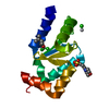
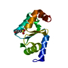
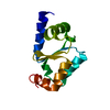
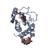
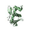
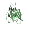
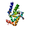
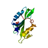
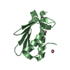
| Title | Crystal structure of cisplatin bound to a plasmodium falciparum glutaredoxin 1 (PfGrx1) | ||||||
 Components Components | Glutaredoxin | ||||||
 Keywords Keywords | OXIDOREDUCTASE / Glutathione / Active site / TRX FOLD / Redox Enzyme / Pt-SAD / cisplatin | ||||||
| Function / homology |  Function and homology informationprotein-disulfide reductase (glutathione) activity / Interconversion of nucleotide di- and triphosphates / glutathione disulfide oxidoreductase activity / antioxidant activity Function and homology informationprotein-disulfide reductase (glutathione) activity / Interconversion of nucleotide di- and triphosphates / glutathione disulfide oxidoreductase activity / antioxidant activitySimilarity search - Function | ||||||
| Biological species |  Plasmodium falciparum (malaria parasite P. falciparum) Plasmodium falciparum (malaria parasite P. falciparum) | ||||||
| Method | X-RAY DIFFRACTION / SAD / Resolution: 1.7 Å | ||||||
 Authors Authors | Yogavel, M. / Sharma, A. | ||||||
 Citation Citation | Journal: To be Published Title: Interaction of Cisplatin with plasmodium falciparum Glutaredoxin 1 Authors: Yogavel, M. / Tripathi, T. / Rahlfs, S. / Becker, K. / Sharma, A. | ||||||
| History |
|
- Structure visualization
| Structure viewer | Molecule: MolmilJmol/JSmol |
|---|
- Downloads & links
-Download
| PDBx/mmCIF format | 4n0z.cif.gz | 35 KB | Display | PDBx/mmCIF format |
|---|---|---|---|---|
| PDB format | pdb4n0z.ent.gz | 26 KB | Display | PDB format |
| PDBx/mmJSON format | 4n0z.json.gz | Tree view | PDBx/mmJSON format | |
| Others |  Other downloads Other downloads |
-Validation report
| Arichive directory | https://data.pdbj.org/pub/pdb/validation_reports/n0/4n0zftp://data.pdbj.org/pub/pdb/validation_reports/n0/4n0z | HTTPS FTP |
|---|
-Related structure data


-Links
 PDBj
PDBj
- Assembly
| Deposited unit | 
| ||||||||
|---|---|---|---|---|---|---|---|---|---|
| 1 |
| ||||||||
| Unit cell |
|
-Components
| #1: Protein | / Glutaredoxin 1 Mass: 12436.457 Da / Num. of mol.: 1 Source method: isolated from a genetically manipulated source Source: (gene. exp.) Plasmodium falciparum (malaria parasite P. falciparum)Strain: 3D7 / Gene: GRX1 / Plasmid: pQE30 / Production host:  Escherichia coli (E. coli) / Strain (production host): M15 Escherichia coli (E. coli) / Strain (production host): M15References: UniProt: Q9NLB2, arsenate reductase (glutathione/glutaredoxin) |
|---|---|
| #2: Chemical | ChemComp-MPO / MOPS  Mass: 209.263 Da / Num. of mol.: 1 / Source method: obtained synthetically / Formula: C7H15NO4S / Comment: pH buffer*YM Mass: 209.263 Da / Num. of mol.: 1 / Source method: obtained synthetically / Formula: C7H15NO4S / Comment: pH buffer*YM |
| #3: Chemical | ChemComp-MPD / (2-Methyl-2,4-pentanediol  Mass: 118.174 Da / Num. of mol.: 1 / Source method: obtained synthetically / Formula: C6H14O2 / Comment: precipitant*YM Mass: 118.174 Da / Num. of mol.: 1 / Source method: obtained synthetically / Formula: C6H14O2 / Comment: precipitant*YM |
| #4: Chemical | ChemComp-CPT / Cisplatin  Mass: 300.045 Da / Num. of mol.: 1 / Source method: obtained synthetically / Formula: Cl2H6N2Pt / Comment: medication, chemotherapy*YM Mass: 300.045 Da / Num. of mol.: 1 / Source method: obtained synthetically / Formula: Cl2H6N2Pt / Comment: medication, chemotherapy*YM |
| #5: Water | ChemComp-HOH / Water Mass: 18.015 Da / Num. of mol.: 107 / Source method: isolated from a natural source / Formula: H2O Mass: 18.015 Da / Num. of mol.: 107 / Source method: isolated from a natural source / Formula: H2O |
-Experimental details
-Experiment
| Experiment | Method: X-RAY DIFFRACTION / Number of used crystals: 1 |
|---|
- Sample preparation
| Crystal | Density Matthews: 2.25 Å3/Da / Density % sol: 45.24 % / Mosaicity: 0.582 ° |
|---|---|
| Crystal grow | Temperature: 293 K / Method: vapor diffusion, hanging drop / pH: 7.5 Details: 12.5%(W/V) PEG1000, 12.5%(W/V) PEG3350, 12.5%(V/V) MPD, 0.02M AMINO ACIDS, 0.1M MOPS/HEPES SODIUM,, pH 7.5, VAPOR DIFFUSION, HANGING DROP, temperature 293K |
-Data collection
| Diffraction | Mean temperature: 100 K | |||||||||||||||||||||||||||||||||||||||||||||||||||||||||||||||||||||||||||||||||||||||||||||||||||||||||||||||||||||||||||||||||||||||||||||||||||
|---|---|---|---|---|---|---|---|---|---|---|---|---|---|---|---|---|---|---|---|---|---|---|---|---|---|---|---|---|---|---|---|---|---|---|---|---|---|---|---|---|---|---|---|---|---|---|---|---|---|---|---|---|---|---|---|---|---|---|---|---|---|---|---|---|---|---|---|---|---|---|---|---|---|---|---|---|---|---|---|---|---|---|---|---|---|---|---|---|---|---|---|---|---|---|---|---|---|---|---|---|---|---|---|---|---|---|---|---|---|---|---|---|---|---|---|---|---|---|---|---|---|---|---|---|---|---|---|---|---|---|---|---|---|---|---|---|---|---|---|---|---|---|---|---|---|---|---|---|
| Diffraction source | Source: ROTATING ANODE / Type: RIGAKU MICROMAX-007 / Wavelength: 1.5418 Å | |||||||||||||||||||||||||||||||||||||||||||||||||||||||||||||||||||||||||||||||||||||||||||||||||||||||||||||||||||||||||||||||||||||||||||||||||||
| Detector | Type: MAR scanner 345 mm plate / Detector: IMAGE PLATE / Date: Jun 26, 2012 / Details: mirrors | |||||||||||||||||||||||||||||||||||||||||||||||||||||||||||||||||||||||||||||||||||||||||||||||||||||||||||||||||||||||||||||||||||||||||||||||||||
| Radiation | Monochromator: SAGITALLY FOCUSED Si(111) / Protocol: SINGLE WAVELENGTH / Monochromatic (M) / Laue (L): M / Scattering type: x-ray | |||||||||||||||||||||||||||||||||||||||||||||||||||||||||||||||||||||||||||||||||||||||||||||||||||||||||||||||||||||||||||||||||||||||||||||||||||
| Radiation wavelength | Wavelength: 1.5418 Å / Relative weight: 1 | |||||||||||||||||||||||||||||||||||||||||||||||||||||||||||||||||||||||||||||||||||||||||||||||||||||||||||||||||||||||||||||||||||||||||||||||||||
| Reflection | Resolution: 1.7→50 Å / Num. all: 12884 / Num. obs: 12755 / % possible obs: 99 % / Observed criterion σ(F): 2 / Observed criterion σ(I): 2 / Redundancy: 22.3 % / Biso Wilson estimate: 22.37 Å2 / Rmerge(I) obs: 0.046 / Χ2: 1.463 / Net I/σ(I): 18.2 | |||||||||||||||||||||||||||||||||||||||||||||||||||||||||||||||||||||||||||||||||||||||||||||||||||||||||||||||||||||||||||||||||||||||||||||||||||
| Reflection shell |
|
- Processing
| Software |
| ||||||||||||||||||||||||||||||||||||
|---|---|---|---|---|---|---|---|---|---|---|---|---|---|---|---|---|---|---|---|---|---|---|---|---|---|---|---|---|---|---|---|---|---|---|---|---|---|
| Refinement | Method to determine structure: SAD / Resolution: 1.7→24.2 Å / Occupancy max: 1 / Occupancy min: 0.26 / FOM work R set: 0.8827 / SU ML: 0.15 / σ(F): 1.36 / Phase error: 17.59 / Stereochemistry target values: ML
| ||||||||||||||||||||||||||||||||||||
| Solvent computation | Shrinkage radii: 0.9 Å / VDW probe radii: 1.11 Å / Solvent model: FLAT BULK SOLVENT MODEL | ||||||||||||||||||||||||||||||||||||
| Displacement parameters | Biso max: 89.73 Å2 / Biso mean: 25.499 Å2 / Biso min: 12.96 Å2 | ||||||||||||||||||||||||||||||||||||
| Refinement step | Cycle: LAST / Resolution: 1.7→24.2 Å
| ||||||||||||||||||||||||||||||||||||
| Refine LS restraints |
| ||||||||||||||||||||||||||||||||||||
| LS refinement shell | Refine-ID: X-RAY DIFFRACTION / Total num. of bins used: 4
|
