 Movie
Movie Controller


+ Open data

- Basic information
| Entry | Database: PDB / ID: 3ctg | ||||||
|---|---|---|---|---|---|---|---|
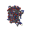
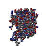
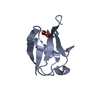
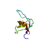
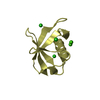
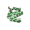
| Title | Crystal structure of reduced glutaredoxin 2 | ||||||
 Components Components | Glutaredoxin-2 | ||||||
 Keywords Keywords | OXIDOREDUCTASE / REDUCED FORM / Electron transport / Mitochondrion / Redox-active center / Transit peptide / Transport | ||||||
| Function / homology |  Function and homology informationglutathione peroxidase / glutathione disulfide oxidoreductase activity / disulfide oxidoreductase activity / glutathione peroxidase activity / glutathione transferase / glutathione transferase activity / glutathione metabolic process / cellular response to oxidative stress / mitochondrion / nucleus ...glutathione peroxidase / glutathione disulfide oxidoreductase activity / disulfide oxidoreductase activity / glutathione peroxidase activity / glutathione transferase / glutathione transferase activity / glutathione metabolic process / cellular response to oxidative stress / mitochondrion / nucleus / cytosol / cytoplasm Function and homology informationglutathione peroxidase / glutathione disulfide oxidoreductase activity / disulfide oxidoreductase activity / glutathione peroxidase activity / glutathione transferase / glutathione transferase activity / glutathione metabolic process / cellular response to oxidative stress / mitochondrion / nucleus ...glutathione peroxidase / glutathione disulfide oxidoreductase activity / disulfide oxidoreductase activity / glutathione peroxidase activity / glutathione transferase / glutathione transferase activity / glutathione metabolic process / cellular response to oxidative stress / mitochondrion / nucleus / cytosol / cytoplasmSimilarity search - Function | ||||||
| Biological species |  Saccharomyces cerevisiae (brewer's yeast) Saccharomyces cerevisiae (brewer's yeast) | ||||||
| Method | X-RAY DIFFRACTION / MOLECULAR REPLACEMENT / Resolution: 1.5 Å | ||||||
 Authors Authors | Yu, J. / Teng, Y.B. / Zhou, C.Z. | ||||||
 Citation Citation | Journal: Biochim.Biophys.Acta / Year: 2010 Title: Structural basis for the different activities of yeast Grx1 and Grx2. Authors: Li, W.F. / Yu, J. / Ma, X.X. / Teng, Y.B. / Luo, M. / Tang, Y.J. / Zhou, C.Z. | ||||||
| History |
|
- Structure visualization
| Structure viewer | Molecule: MolmilJmol/JSmol |
|---|
- Downloads & links
-Download
| PDBx/mmCIF format | 3ctg.cif.gz | 39 KB | Display | PDBx/mmCIF format |
|---|---|---|---|---|
| PDB format | pdb3ctg.ent.gz | 25.2 KB | Display | PDB format |
| PDBx/mmJSON format | 3ctg.json.gz | Tree view | PDBx/mmJSON format | |
| Others |  Other downloads Other downloads |
-Validation report
| Arichive directory | https://data.pdbj.org/pub/pdb/validation_reports/ct/3ctgftp://data.pdbj.org/pub/pdb/validation_reports/ct/3ctg | HTTPS FTP |
|---|
-Related structure data
| Related structure data |  3ctfSC S: Starting model for refinement C: citing same article ( |
|---|---|
| Similar structure data |


-Links
 PDBj
PDBj


- Assembly
| Deposited unit | 
| ||||||||
|---|---|---|---|---|---|---|---|---|---|
| 1 |
| ||||||||
| Unit cell |
|
-Components
| #1: Protein | / Thioltransferase / Glutathione-dependent oxidoreductase 2 Mass: 14150.113 Da / Num. of mol.: 1 Source method: isolated from a genetically manipulated source Source: (gene. exp.) Saccharomyces cerevisiae (brewer's yeast)Strain: S288C / Gene: GRX2, TTR, TTR1, YDR513W, D9719.17 / Plasmid: PET15B / Production host:  Escherichia coli (E. coli) / Strain (production host): BL21(DE3) Escherichia coli (E. coli) / Strain (production host): BL21(DE3)References: UniProt: P17695, arsenate reductase (glutathione/glutaredoxin) |
|---|---|
| #2: Water | ChemComp-HOH / Water Mass: 18.015 Da / Num. of mol.: 211 / Source method: isolated from a natural source / Formula: H2O Mass: 18.015 Da / Num. of mol.: 211 / Source method: isolated from a natural source / Formula: H2O |
-Experimental details
-Experiment
| Experiment | Method: X-RAY DIFFRACTION / Number of used crystals: 1 |
|---|
- Sample preparation
| Crystal | Density Matthews: 2.4 Å3/Da / Density % sol: 48.74 % |
|---|---|
| Crystal grow | Temperature: 289 K / Method: vapor diffusion, hanging drop / pH: 4.6 Details: 30% PEG 4000, 0.1M NAAC, 0.2M NH4AC, VAPOR DIFFUSION, HANGING DROP, pH 4.60, temperature 289K |
-Data collection
| Diffraction | Mean temperature: 100 K |
|---|---|
| Diffraction source | Source: ROTATING ANODE / Type: RIGAKU / Wavelength: 1.54179 Å |
| Detector | Type: MAR scanner 345 mm plate / Detector: IMAGE PLATE / Details: MIRRORS |
| Radiation | Monochromator: YALE MIRRORS / Protocol: SINGLE WAVELENGTH / Monochromatic (M) / Laue (L): M / Scattering type: x-ray |
| Radiation wavelength | Wavelength: 1.54179 Å / Relative weight: 1 |
| Reflection | Resolution: 1.5→22.13 Å / Num. obs: 20618 / % possible obs: 98.5 % / Redundancy: 2.91 % / Biso Wilson estimate: 16.5 Å2 / Rmerge(I) obs: 0.076 / Net I/σ(I): 6.4 |
| Reflection shell | Resolution: 1.5→1.55 Å / Redundancy: 2.97 % / Rmerge(I) obs: 0.141 / Mean I/σ(I) obs: 3.3 / % possible all: 99.4 |
- Processing
| Software |
| ||||||||||||||||||||||||||||||||||||||||||||||||||||||||||||
|---|---|---|---|---|---|---|---|---|---|---|---|---|---|---|---|---|---|---|---|---|---|---|---|---|---|---|---|---|---|---|---|---|---|---|---|---|---|---|---|---|---|---|---|---|---|---|---|---|---|---|---|---|---|---|---|---|---|---|---|---|---|
| Refinement | Method to determine structure: MOLECULAR REPLACEMENT Starting model: PDB ENTRY 3CTF Resolution: 1.5→22.13 Å / Rfactor Rfree error: 0.006 / Data cutoff high absF: 727971.05 / Data cutoff low absF: 0 / Isotropic thermal model: RESTRAINED / Cross valid method: THROUGHOUT / σ(F): 0 / Stereochemistry target values: Engh & Huber
| ||||||||||||||||||||||||||||||||||||||||||||||||||||||||||||
| Solvent computation | Solvent model: FLAT MODEL / Bsol: 45.73 Å2 / ksol: 0.39 e/Å3 | ||||||||||||||||||||||||||||||||||||||||||||||||||||||||||||
| Displacement parameters | Biso mean: 17.1 Å2
| ||||||||||||||||||||||||||||||||||||||||||||||||||||||||||||
| Refine analyze |
| ||||||||||||||||||||||||||||||||||||||||||||||||||||||||||||
| Refinement step | Cycle: LAST / Resolution: 1.5→22.13 Å
| ||||||||||||||||||||||||||||||||||||||||||||||||||||||||||||
| Refine LS restraints |
| ||||||||||||||||||||||||||||||||||||||||||||||||||||||||||||
| LS refinement shell | Resolution: 1.5→1.55 Å / Rfactor Rfree error: 0.029 / Total num. of bins used: 10
| ||||||||||||||||||||||||||||||||||||||||||||||||||||||||||||
| Xplor file |
|
