 Movie
Movie Controller


+ Open data

- Basic information
| Entry | Database: PDB / ID: 4mwi | ||||||
|---|---|---|---|---|---|---|---|
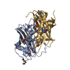
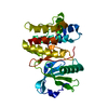
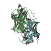
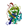
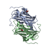
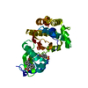
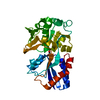
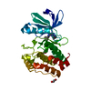
| Title | Crystal structure of the human MLKL pseudokinase domain | ||||||
 Components Components | Mixed lineage kinase domain-like protein | ||||||
 Keywords Keywords |  TRANSFERASE / Pseudokinase / necroptosis TRANSFERASE / Pseudokinase / necroptosis | ||||||
| Function / homology |  Function and homology information Function and homology informationexecution phase of necroptosis / Microbial modulation of RIPK1-mediated regulated necrosis / necroptotic signaling pathway / TRP channels / RIPK1-mediated regulated necrosis / protein homotrimerization / necroptotic process / Regulation of necroptotic cell death / cell junction / defense response to virus ...execution phase of necroptosis / Microbial modulation of RIPK1-mediated regulated necrosis / necroptotic signaling pathway / TRP channels / RIPK1-mediated regulated necrosis / protein homotrimerization / necroptotic process / Regulation of necroptotic cell death / cell junction / defense response to virus / cell surface receptor signaling pathway / protein-containing complex binding / protein kinase binding / ATP binding / identical protein binding / nucleus / plasma membrane / cytosol / cytoplasmSimilarity search - Function | ||||||
| Biological species |  Homo sapiens (human) Homo sapiens (human) | ||||||
| Method | X-RAY DIFFRACTION / SYNCHROTRON / MOLECULAR REPLACEMENT / Resolution: 1.7 Å | ||||||
 Authors Authors | Czabotar, P.E. / Murphy, J.M. | ||||||
 Citation Citation | Journal: Biochem.J. / Year: 2014 Title: Insights into the evolution of divergent nucleotide-binding mechanisms among pseudokinases revealed by crystal structures of human and mouse MLKL. Authors: Murphy, J.M. / Lucet, I.S. / Hildebrand, J.M. / Tanzer, M.C. / Young, S.N. / Sharma, P. / Lessene, G. / Alexander, W.S. / Babon, J.J. / Silke, J. / Czabotar, P.E. | ||||||
| History |
|
- Structure visualization
| Structure viewer | Molecule: MolmilJmol/JSmol |
|---|
- Downloads & links
-Download
| PDBx/mmCIF format | 4mwi.cif.gz | 131.1 KB | Display | PDBx/mmCIF format |
|---|---|---|---|---|
| PDB format | pdb4mwi.ent.gz | 100.8 KB | Display | PDB format |
| PDBx/mmJSON format | 4mwi.json.gz | Tree view | PDBx/mmJSON format | |
| Others |  Other downloads Other downloads |
-Validation report
| Arichive directory | https://data.pdbj.org/pub/pdb/validation_reports/mw/4mwiftp://data.pdbj.org/pub/pdb/validation_reports/mw/4mwi | HTTPS FTP |
|---|
-Related structure data
| Related structure data | 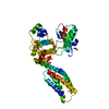 4btfS S: Starting model for refinement |
|---|---|
| Similar structure data |


-Links
 PDBj
PDBj

- Assembly
| Deposited unit | 
| |||||||||
|---|---|---|---|---|---|---|---|---|---|---|
| 1 |
| |||||||||
| Unit cell |
| |||||||||
| Components on special symmetry positions |
|
-Components
| #1: Protein | Mass: 33567.789 Da / Num. of mol.: 1 / Fragment: unp residues 183-471 Source method: isolated from a genetically manipulated source Source: (gene. exp.) Homo sapiens (human) / Gene: MLKL / Production host:   Spodoptera frugiperda (fall armyworm) / References: UniProt: Q8NB16 Spodoptera frugiperda (fall armyworm) / References: UniProt: Q8NB16 |
|---|---|
| #2: Chemical | ChemComp-PXN / (  Mass: 368.463 Da / Num. of mol.: 1 / Source method: obtained synthetically / Formula: C17H36O8 Mass: 368.463 Da / Num. of mol.: 1 / Source method: obtained synthetically / Formula: C17H36O8 |
| #3: Chemical | ChemComp-GOL / Glycerol  Mass: 92.094 Da / Num. of mol.: 1 / Source method: obtained synthetically / Formula: C3H8O3 Mass: 92.094 Da / Num. of mol.: 1 / Source method: obtained synthetically / Formula: C3H8O3 |
| #4: Water | ChemComp-HOH / Water Mass: 18.015 Da / Num. of mol.: 325 / Source method: isolated from a natural source / Formula: H2O Mass: 18.015 Da / Num. of mol.: 325 / Source method: isolated from a natural source / Formula: H2O |
-Experimental details
-Experiment
| Experiment | Method: X-RAY DIFFRACTION / Number of used crystals: 1 |
|---|
- Sample preparation
| Crystal | Density Matthews: 2.56 Å3/Da / Density % sol: 51.96 % |
|---|---|
| Crystal grow | Temperature: 293 K / Method: vapor diffusion, sitting drop / pH: 8.5 Details: 25% w/v pentaerythitol propoxylate, 0.1M Tris chloride, pH 8.5, VAPOR DIFFUSION, SITTING DROP, temperature 293K |
-Data collection
| Diffraction | Mean temperature: 100 K |
|---|---|
| Diffraction source | Source: SYNCHROTRON / Site: Australian Synchrotron  / Beamline: MX2 / Wavelength: 0.9537 / Beamline: MX2 / Wavelength: 0.9537 |
| Detector | Type: ADSC QUANTUM 315r / Detector: CCD / Date: Apr 9, 2013 |
| Radiation | Monochromator: SI 111 CHANNEL / Protocol: SINGLE WAVELENGTH / Monochromatic (M) / Laue (L): M / Scattering type: x-ray |
| Radiation wavelength | Wavelength: 0.9537 Å / Relative weight: 1 |
| Reflection | Resolution: 1.7→50 Å / Num. obs: 38267 / % possible obs: 98.9 % / Observed criterion σ(I): -3 / Redundancy: 7.04 % / Rmerge(I) obs: 0.057 / Net I/σ(I): 21.87 |
| Reflection shell | Resolution: 1.7→1.8 Å / Redundancy: 5.74 % / Rmerge(I) obs: 0.884 / Mean I/σ(I) obs: 1.94 / % possible all: 99.7 |
- Processing
| Software |
| |||||||||||||||||||||||||||||||||||||||||||||||||||||||||||||||||||||||||||||||||||||||||||||||||||||||||
|---|---|---|---|---|---|---|---|---|---|---|---|---|---|---|---|---|---|---|---|---|---|---|---|---|---|---|---|---|---|---|---|---|---|---|---|---|---|---|---|---|---|---|---|---|---|---|---|---|---|---|---|---|---|---|---|---|---|---|---|---|---|---|---|---|---|---|---|---|---|---|---|---|---|---|---|---|---|---|---|---|---|---|---|---|---|---|---|---|---|---|---|---|---|---|---|---|---|---|---|---|---|---|---|---|---|---|
| Refinement | Method to determine structure: MOLECULAR REPLACEMENT Starting model: PDB ENTRY 4BTF Resolution: 1.7→19.68 Å / SU ML: 0.19 / σ(F): 2 / Phase error: 18.36 / Stereochemistry target values: ML
| |||||||||||||||||||||||||||||||||||||||||||||||||||||||||||||||||||||||||||||||||||||||||||||||||||||||||
| Solvent computation | Shrinkage radii: 0.9 Å / VDW probe radii: 1.11 Å / Solvent model: FLAT BULK SOLVENT MODEL | |||||||||||||||||||||||||||||||||||||||||||||||||||||||||||||||||||||||||||||||||||||||||||||||||||||||||
| Refinement step | Cycle: LAST / Resolution: 1.7→19.68 Å
| |||||||||||||||||||||||||||||||||||||||||||||||||||||||||||||||||||||||||||||||||||||||||||||||||||||||||
| Refine LS restraints |
| |||||||||||||||||||||||||||||||||||||||||||||||||||||||||||||||||||||||||||||||||||||||||||||||||||||||||
| LS refinement shell |
| |||||||||||||||||||||||||||||||||||||||||||||||||||||||||||||||||||||||||||||||||||||||||||||||||||||||||
| Refinement TLS params. | Method: refined / Refine-ID: X-RAY DIFFRACTION
| |||||||||||||||||||||||||||||||||||||||||||||||||||||||||||||||||||||||||||||||||||||||||||||||||||||||||
| Refinement TLS group |
|
