| Entry | Database: PDB / ID: 4mrc
|
|---|
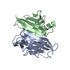
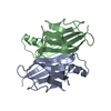
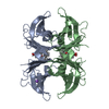
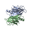
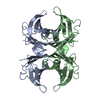
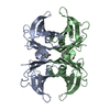
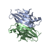
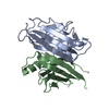
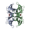
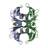
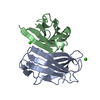
| Title | Human Transthyretin Ser52Pro Mutant |
|---|
 Components Components | Transthyretin |
|---|
 Keywords Keywords | TRANSPORT PROTEIN / Hormone Transporter / Thyroxine T4 |
|---|
| Function / homology |  Function and homology information Function and homology information |
|---|
| Biological species |  Homo sapiens (human) Homo sapiens (human) |
|---|
| Method | X-RAY DIFFRACTION / SYNCHROTRON / MOLECULAR REPLACEMENT / Resolution: 1.54 Å |
|---|
 Authors Authors | Chen, W.J. / Wood, S.P. |
|---|
 Citation Citation | Journal: Proc.Natl.Acad.Sci.USA / Year: 2014
Title: Proteolytic cleavage of Ser52Pro variant transthyretin triggers its amyloid fibrillogenesis.
Authors: Mangione, P.P. / Porcari, R. / Gillmore, J.D. / Pucci, P. / Monti, M. / Porcari, M. / Giorgetti, S. / Marchese, L. / Raimondi, S. / Serpell, L.C. / Chen, W. / Relini, A. / Marcoux, J. / ...Authors: Mangione, P.P. / Porcari, R. / Gillmore, J.D. / Pucci, P. / Monti, M. / Porcari, M. / Giorgetti, S. / Marchese, L. / Raimondi, S. / Serpell, L.C. / Chen, W. / Relini, A. / Marcoux, J. / Clatworthy, I.R. / Taylor, G.W. / Tennent, G.A. / Robinson, C.V. / Hawkins, P.N. / Stoppini, M. / Wood, S.P. / Pepys, M.B. / Bellotti, V. |
|---|
| History | | Deposition | Sep 17, 2013 | Deposition site: RCSB / Processing site: RCSB |
|---|
| Revision 1.0 | Jan 8, 2014 | Provider: repository / Type: Initial release |
|---|
| Revision 1.1 | Jan 15, 2014 | Group: Database references |
|---|
| Revision 1.2 | Feb 26, 2014 | Group: Database references |
|---|
| Revision 1.3 | Feb 28, 2024 | Group: Data collection / Database references / Derived calculations
Category: chem_comp_atom / chem_comp_bond ...chem_comp_atom / chem_comp_bond / database_2 / pdbx_struct_conn_angle / struct_conn / struct_ref_seq_dif / struct_site
Item: _database_2.pdbx_DOI / _database_2.pdbx_database_accession ..._database_2.pdbx_DOI / _database_2.pdbx_database_accession / _pdbx_struct_conn_angle.ptnr1_auth_seq_id / _pdbx_struct_conn_angle.ptnr3_auth_seq_id / _pdbx_struct_conn_angle.value / _struct_conn.pdbx_dist_value / _struct_conn.ptnr1_auth_asym_id / _struct_conn.ptnr1_auth_comp_id / _struct_conn.ptnr1_auth_seq_id / _struct_conn.ptnr1_label_asym_id / _struct_conn.ptnr1_label_atom_id / _struct_conn.ptnr1_label_comp_id / _struct_conn.ptnr1_label_seq_id / _struct_conn.ptnr2_auth_asym_id / _struct_conn.ptnr2_auth_comp_id / _struct_conn.ptnr2_auth_seq_id / _struct_conn.ptnr2_label_asym_id / _struct_conn.ptnr2_label_atom_id / _struct_conn.ptnr2_label_comp_id / _struct_ref_seq_dif.details / _struct_site.pdbx_auth_asym_id / _struct_site.pdbx_auth_comp_id / _struct_site.pdbx_auth_seq_id |
|---|
|
|---|
 Movie
Movie Controller


 Open data
 Basic information
 Structure visualization
 Downloads & links
 Other downloads
 PDBj
PDBj


 Assembly


 Mass: 40.078 Da / Num. of mol.: 2 / Source method: obtained synthetically / Formula: Ca
 Mass: 18.015 Da / Num. of mol.: 88 / Source method: isolated from a natural source / Formula: H2O
 Sample preparation
 / Beamline: I04 / Wavelength: 0.979 Å
 Processing