 Movie
Movie Controller


+ Open data

- Basic information
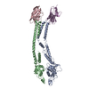
| Entry | Database: PDB / ID: 4m4p | |||||||||
|---|---|---|---|---|---|---|---|---|---|---|
| Title | Crystal structure of EPHA4 ectodomain | |||||||||
 Components Components | Ephrin type-A receptor 4 | |||||||||
 Keywords Keywords |  TRANSFERASE / eph receptor protein kinase TRANSFERASE / eph receptor protein kinase | |||||||||
| Function / homology |  Function and homology informationDH domain binding / neuron projection fasciculation / positive regulation of Rho guanyl-nucleotide exchange factor activity / corticospinal tract morphogenesis / regulation of astrocyte differentiation / negative regulation of proteolysis involved in protein catabolic process / neuron projection guidance / nephric duct morphogenesis / regulation of synapse pruning / fasciculation of sensory neuron axon ...DH domain binding / neuron projection fasciculation / positive regulation of Rho guanyl-nucleotide exchange factor activity / corticospinal tract morphogenesis / regulation of astrocyte differentiation / negative regulation of proteolysis involved in protein catabolic process / neuron projection guidance / nephric duct morphogenesis / regulation of synapse pruning / fasciculation of sensory neuron axon / fasciculation of motor neuron axon / synapse pruning / positive regulation of aspartic-type endopeptidase activity involved in amyloid precursor protein catabolic process / negative regulation of cellular response to hypoxia / negative regulation of axon regeneration / glial cell migration / PH domain binding / GPI-linked ephrin receptor activity / regulation of modification of synaptic structure / transmembrane-ephrin receptor activity / regulation of dendritic spine morphogenesis / negative regulation of epithelial to mesenchymal transition / negative regulation of cell adhesion / motor neuron axon guidance / adult walking behavior / adherens junction organization / positive regulation of dendrite morphogenesis / EPH-Ephrin signaling / Somitogenesis / positive regulation of amyloid-beta formation / regulation of axonogenesis / cochlea development / regulation of GTPase activity / EPHA-mediated growth cone collapse / positive regulation of cell adhesion / positive regulation of protein tyrosine kinase activity / negative regulation of long-term synaptic potentiation / EPH-ephrin mediated repulsion of cells / ephrin receptor signaling pathway / axon terminus / axonal growth cone / ephrin receptor binding / negative regulation of cell migration / protein tyrosine kinase binding / dendritic shaft / filopodium / axon guidance / postsynaptic density membrane / adherens junction / Schaffer collateral - CA1 synapse / neuromuscular junction / negative regulation of ERK1 and ERK2 cascade / receptor protein-tyrosine kinase / peptidyl-tyrosine phosphorylation / cellular response to amyloid-beta / negative regulation of neuron projection development / presynaptic membrane / kinase activity / amyloid-beta binding / early endosome membrane / perikaryon / protein tyrosine kinase activity / mitochondrial outer membrane / dendritic spine / protein autophosphorylation / negative regulation of translation / protein stabilization / cell adhesion / protein kinase activity / positive regulation of cell migration / axon / glutamatergic synapse / dendrite / positive regulation of cell population proliferation / cell surface / ATP binding / identical protein binding / plasma membrane / cytoplasm Function and homology informationDH domain binding / neuron projection fasciculation / positive regulation of Rho guanyl-nucleotide exchange factor activity / corticospinal tract morphogenesis / regulation of astrocyte differentiation / negative regulation of proteolysis involved in protein catabolic process / neuron projection guidance / nephric duct morphogenesis / regulation of synapse pruning / fasciculation of sensory neuron axon ...DH domain binding / neuron projection fasciculation / positive regulation of Rho guanyl-nucleotide exchange factor activity / corticospinal tract morphogenesis / regulation of astrocyte differentiation / negative regulation of proteolysis involved in protein catabolic process / neuron projection guidance / nephric duct morphogenesis / regulation of synapse pruning / fasciculation of sensory neuron axon / fasciculation of motor neuron axon / synapse pruning / positive regulation of aspartic-type endopeptidase activity involved in amyloid precursor protein catabolic process / negative regulation of cellular response to hypoxia / negative regulation of axon regeneration / glial cell migration / PH domain binding / GPI-linked ephrin receptor activity / regulation of modification of synaptic structure / transmembrane-ephrin receptor activity / regulation of dendritic spine morphogenesis / negative regulation of epithelial to mesenchymal transition / negative regulation of cell adhesion / motor neuron axon guidance / adult walking behavior / adherens junction organization / positive regulation of dendrite morphogenesis / EPH-Ephrin signaling / Somitogenesis / positive regulation of amyloid-beta formation / regulation of axonogenesis / cochlea development / regulation of GTPase activity / EPHA-mediated growth cone collapse / positive regulation of cell adhesion / positive regulation of protein tyrosine kinase activity / negative regulation of long-term synaptic potentiation / EPH-ephrin mediated repulsion of cells / ephrin receptor signaling pathway / axon terminus / axonal growth cone / ephrin receptor binding / negative regulation of cell migration / protein tyrosine kinase binding / dendritic shaft / filopodium / axon guidance / postsynaptic density membrane / adherens junction / Schaffer collateral - CA1 synapse / neuromuscular junction / negative regulation of ERK1 and ERK2 cascade / receptor protein-tyrosine kinase / peptidyl-tyrosine phosphorylation / cellular response to amyloid-beta / negative regulation of neuron projection development / presynaptic membrane / kinase activity / amyloid-beta binding / early endosome membrane / perikaryon / protein tyrosine kinase activity / mitochondrial outer membrane / dendritic spine / protein autophosphorylation / negative regulation of translation / protein stabilization / cell adhesion / protein kinase activity / positive regulation of cell migration / axon / glutamatergic synapse / dendrite / positive regulation of cell population proliferation / cell surface / ATP binding / identical protein binding / plasma membrane / cytoplasmSimilarity search - Function | |||||||||
| Biological species |  Homo sapiens (human) Homo sapiens (human) | |||||||||
| Method | X-RAY DIFFRACTION / SYNCHROTRON / MOLECULAR REPLACEMENT / Resolution: 2.081 Å | |||||||||
 Authors Authors | Xu, K. / Tsvetkova-Robev, D. / Xu, Y. / Goldgur, Y. / Chan, Y.-P. / Himanen, J.P. / Nikolov, D.B. | |||||||||
 Citation Citation | Journal: Proc.Natl.Acad.Sci.USA / Year: 2013 Title: Insights into Eph receptor tyrosine kinase activation from crystal structures of the EphA4 ectodomain and its complex with ephrin-A5. Authors: Xu, K. / Tzvetkova-Robev, D. / Xu, Y. / Goldgur, Y. / Chan, Y.P. / Himanen, J.P. / Nikolov, D.B. | |||||||||
| History |
|
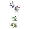
- Structure visualization
| Structure viewer | Molecule: MolmilJmol/JSmol |
|---|
- Downloads & links
-Download
| PDBx/mmCIF format | 4m4p.cif.gz | 223.3 KB | Display | PDBx/mmCIF format |
|---|---|---|---|---|
| PDB format | pdb4m4p.ent.gz | 177.2 KB | Display | PDB format |
| PDBx/mmJSON format | 4m4p.json.gz | Tree view | PDBx/mmJSON format | |
| Others |  Other downloads Other downloads |
-Validation report
| Arichive directory | https://data.pdbj.org/pub/pdb/validation_reports/m4/4m4pftp://data.pdbj.org/pub/pdb/validation_reports/m4/4m4p | HTTPS FTP |
|---|
-Related structure data
| Related structure data |  4m4rC  3fl7S C: citing same article ( S: Starting model for refinement |
|---|---|
| Similar structure data |


-Links
 PDBj
PDBj


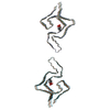
- Assembly
| Deposited unit | 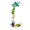
| ||||||||
|---|---|---|---|---|---|---|---|---|---|
| 1 |
| ||||||||
| Unit cell |
|
-Components
| #1: Protein | Mass: 57414.156 Da / Num. of mol.: 1 / Fragment: UNP residues 27-543 Source method: isolated from a genetically manipulated source Source: (gene. exp.) Homo sapiens (human) / Gene: EPHA4, HEK8, SEK, TYRO1 / Organ (production host): ovary / Production host:  Trichoplusia ni (cabbage looper) / Strain (production host): high5 Trichoplusia ni (cabbage looper) / Strain (production host): high5References: UniProt: P54764, receptor protein-tyrosine kinase | ||
|---|---|---|---|
| #2: Polysaccharide | 2-acetamido-2-deoxy-beta-D-glucopyranose-(1-4)-2-acetamido-2-deoxy-beta-D-glucopyranose / Mass: 424.401 Da / Num. of mol.: 1 Source method: isolated from a genetically manipulated source | ||
| #3: Sugar | N-Acetylglucosamine  Type: D-saccharide, beta linking / Mass: 221.208 Da / Num. of mol.: 2 Type: D-saccharide, beta linking / Mass: 221.208 Da / Num. of mol.: 2Source method: isolated from a genetically manipulated source Formula: C8H15NO6 #4: Water | ChemComp-HOH / | Water Mass: 18.015 Da / Num. of mol.: 161 / Source method: isolated from a natural source / Formula: H2O Mass: 18.015 Da / Num. of mol.: 161 / Source method: isolated from a natural source / Formula: H2O |
-Experimental details
-Experiment
| Experiment | Method: X-RAY DIFFRACTION / Number of used crystals: 1 |
|---|
- Sample preparation
| Crystal | Density Matthews: 3.16 Å3/Da / Density % sol: 61.05 % |
|---|---|
| Crystal grow | Temperature: 298 K / Method: vapor diffusion, sitting drop / pH: 6 Details: 22% PEG400, 0.1M MES buffer pH 6.0, 3% DMSO, VAPOR DIFFUSION, SITTING DROP, temperature 298K |
-Data collection
| Diffraction | Mean temperature: 100 K |
|---|---|
| Diffraction source | Source: SYNCHROTRON / Site: APS  / Beamline: 24-ID-C / Wavelength: 0.9792 Å / Beamline: 24-ID-C / Wavelength: 0.9792 Å |
| Detector | Type: ADSC QUANTUM 315 / Detector: CCD / Date: Feb 15, 2011 |
| Radiation | Protocol: SINGLE WAVELENGTH / Monochromatic (M) / Laue (L): M / Scattering type: x-ray |
| Radiation wavelength | Wavelength: 0.9792 Å / Relative weight: 1 |
| Reflection | Resolution: 2.08→27 Å / Num. all: 42116 / Num. obs: 42061 / % possible obs: 99.87 % / Observed criterion σ(F): 2 / Observed criterion σ(I): -3 |
| Reflection shell | Resolution: 2.08→2.14 Å / Redundancy: 3.8 % / Rmerge(I) obs: 0.68 / Mean I/σ(I) obs: 2.2 / % possible all: 100 |
- Processing
| Software |
| ||||||||||||||||||||||||||||||||||||||||||||||||||||||||||||||||||||||||||||||||||||||||||||||||||||||||||||||||||||||||||||||||||||||||||||||||||||||
|---|---|---|---|---|---|---|---|---|---|---|---|---|---|---|---|---|---|---|---|---|---|---|---|---|---|---|---|---|---|---|---|---|---|---|---|---|---|---|---|---|---|---|---|---|---|---|---|---|---|---|---|---|---|---|---|---|---|---|---|---|---|---|---|---|---|---|---|---|---|---|---|---|---|---|---|---|---|---|---|---|---|---|---|---|---|---|---|---|---|---|---|---|---|---|---|---|---|---|---|---|---|---|---|---|---|---|---|---|---|---|---|---|---|---|---|---|---|---|---|---|---|---|---|---|---|---|---|---|---|---|---|---|---|---|---|---|---|---|---|---|---|---|---|---|---|---|---|---|---|---|---|
| Refinement | Method to determine structure: MOLECULAR REPLACEMENT Starting model: 3fl7 Resolution: 2.081→27 Å / SU ML: 0.22 / Cross valid method: THROUGHOUT / σ(F): 1.99 / Phase error: 22.6 / Stereochemistry target values: ML Details: THE AUTHORS STATE THAT THE CONFORMATION ISSUE FOR NAG601A AS WELL AS THE HIGH REAL SPACE R-FACTORS FOR NAG601A AND NAG602A ARE DUE TO THE PARTIAL DISORDER. THE DENSITY IS OF POOR QUALITY AND ...Details: THE AUTHORS STATE THAT THE CONFORMATION ISSUE FOR NAG601A AS WELL AS THE HIGH REAL SPACE R-FACTORS FOR NAG601A AND NAG602A ARE DUE TO THE PARTIAL DISORDER. THE DENSITY IS OF POOR QUALITY AND THE MODEL PRIMARILY REFLECTS THAT THEY ARE THERE.
| ||||||||||||||||||||||||||||||||||||||||||||||||||||||||||||||||||||||||||||||||||||||||||||||||||||||||||||||||||||||||||||||||||||||||||||||||||||||
| Solvent computation | Shrinkage radii: 0.9 Å / VDW probe radii: 1.11 Å / Solvent model: FLAT BULK SOLVENT MODEL | ||||||||||||||||||||||||||||||||||||||||||||||||||||||||||||||||||||||||||||||||||||||||||||||||||||||||||||||||||||||||||||||||||||||||||||||||||||||
| Refinement step | Cycle: LAST / Resolution: 2.081→27 Å
| ||||||||||||||||||||||||||||||||||||||||||||||||||||||||||||||||||||||||||||||||||||||||||||||||||||||||||||||||||||||||||||||||||||||||||||||||||||||
| Refine LS restraints |
| ||||||||||||||||||||||||||||||||||||||||||||||||||||||||||||||||||||||||||||||||||||||||||||||||||||||||||||||||||||||||||||||||||||||||||||||||||||||
| LS refinement shell |
| ||||||||||||||||||||||||||||||||||||||||||||||||||||||||||||||||||||||||||||||||||||||||||||||||||||||||||||||||||||||||||||||||||||||||||||||||||||||
| Refinement TLS params. | Method: refined / Refine-ID: X-RAY DIFFRACTION
| ||||||||||||||||||||||||||||||||||||||||||||||||||||||||||||||||||||||||||||||||||||||||||||||||||||||||||||||||||||||||||||||||||||||||||||||||||||||
| Refinement TLS group |
|
