[English] 日本語
Yorodumi
- PDB-4bk4: crystal structure of the human EphA4 ectodomain -

+
Open data


ID or keywords:

Loading...

-
Basic information

Entry
Database: PDB / ID: 4bk4
Titlecrystal structure of the human EphA4 ectodomain
ComponentsEPHRIN TYPE-A RECEPTOR 4
KeywordsSIGNALING PROTEIN / CELL ADHESION / CELL REPULSION / RECEPTOR CLUSTERING / RECEPTOR CIS INTERACTION / EPH-EPHRIN CRYSTAL STRUCTURES / EPH ECTODOMAIN / ERYTHROPOETIN-PRODUCING HEPATOCELLULAR RECEPTOR / LBD / EGF / FN
Function / homology
Function and homology information


DH domain binding / neuron projection fasciculation / positive regulation of Rho guanyl-nucleotide exchange factor activity / corticospinal tract morphogenesis / regulation of astrocyte differentiation / negative regulation of proteolysis involved in protein catabolic process / neuron projection guidance / nephric duct morphogenesis / regulation of synapse pruning / fasciculation of sensory neuron axon ...DH domain binding / neuron projection fasciculation / positive regulation of Rho guanyl-nucleotide exchange factor activity / corticospinal tract morphogenesis / regulation of astrocyte differentiation / negative regulation of proteolysis involved in protein catabolic process / neuron projection guidance / nephric duct morphogenesis / regulation of synapse pruning / fasciculation of sensory neuron axon / fasciculation of motor neuron axon / synapse pruning / negative regulation of cellular response to hypoxia / negative regulation of axon regeneration / positive regulation of aspartic-type endopeptidase activity involved in amyloid precursor protein catabolic process / glial cell migration / transmembrane-ephrin receptor activity / PH domain binding / GPI-linked ephrin receptor activity / regulation of modification of synaptic structure / negative regulation of epithelial to mesenchymal transition / regulation of dendritic spine morphogenesis / negative regulation of cell adhesion / motor neuron axon guidance / adult walking behavior / adherens junction organization / positive regulation of dendrite morphogenesis / EPH-Ephrin signaling / Somitogenesis / regulation of axonogenesis / positive regulation of amyloid-beta formation / cochlea development / regulation of GTPase activity / EPHA-mediated growth cone collapse / negative regulation of long-term synaptic potentiation / positive regulation of cell adhesion / EPH-ephrin mediated repulsion of cells / ephrin receptor signaling pathway / positive regulation of protein tyrosine kinase activity / axon terminus / axonal growth cone / protein tyrosine kinase binding / ephrin receptor binding / negative regulation of cell migration / dendritic shaft / filopodium / axon guidance / postsynaptic density membrane / adherens junction / Schaffer collateral - CA1 synapse / neuromuscular junction / negative regulation of ERK1 and ERK2 cascade / receptor protein-tyrosine kinase / peptidyl-tyrosine phosphorylation / cellular response to amyloid-beta / negative regulation of neuron projection development / presynaptic membrane / amyloid-beta binding / kinase activity / perikaryon / early endosome membrane / protein tyrosine kinase activity / mitochondrial outer membrane / dendritic spine / protein autophosphorylation / negative regulation of translation / protein stabilization / cell adhesion / protein kinase activity / positive regulation of cell migration / axon / glutamatergic synapse / dendrite / positive regulation of cell population proliferation / cell surface / ATP binding / identical protein binding / plasma membrane / cytoplasm
Similarity search - Function
ephrin a2 ectodomain / Ephrin type-A receptor 4, SAM domain / Ephrin type-A receptor 4, ligand binding domain / Tumor Necrosis Factor Receptor, subunit A, domain 2 / Tumor Necrosis Factor Receptor, subunit A; domain 2 / Tyrosine-protein kinase ephrin type A/B receptor-like / Tyrosine-protein kinase ephrin type A/B receptor-like / Ephrin receptor type-A /type-B / Ephrin receptor ligand binding domain / Tyrosine-protein kinase, receptor class V, conserved site ...ephrin a2 ectodomain / Ephrin type-A receptor 4, SAM domain / Ephrin type-A receptor 4, ligand binding domain / Tumor Necrosis Factor Receptor, subunit A, domain 2 / Tumor Necrosis Factor Receptor, subunit A; domain 2 / Tyrosine-protein kinase ephrin type A/B receptor-like / Tyrosine-protein kinase ephrin type A/B receptor-like / Ephrin receptor type-A /type-B / Ephrin receptor ligand binding domain / Tyrosine-protein kinase, receptor class V, conserved site / Ephrin receptor, transmembrane domain / Ephrin receptor ligand binding domain / Ephrin type-A receptor 2 transmembrane domain / Receptor tyrosine kinase class V signature 1. / Receptor tyrosine kinase class V signature 2. / Eph receptor ligand-binding domain profile. / Ephrin receptor ligand binding domain / Putative ephrin-receptor like / SAM domain (Sterile alpha motif) / Galactose-binding domain-like / SAM domain profile. / Sterile alpha motif. / Sterile alpha motif domain / Sterile alpha motif/pointed domain superfamily / Fibronectin type III domain / Fibronectin type 3 domain / Fibronectin type-III domain profile. / Galactose-binding-like domain superfamily / Fibronectin type III / Fibronectin type III superfamily / Ribbon / Tyrosine-protein kinase, catalytic domain / Tyrosine kinase, catalytic domain / Tyrosine protein kinases specific active-site signature. / Tyrosine-protein kinase, active site / Protein tyrosine and serine/threonine kinase / Serine-threonine/tyrosine-protein kinase, catalytic domain / Jelly Rolls / Serine/Threonine protein kinases, catalytic domain / Immunoglobulins / Protein kinase, ATP binding site / Protein kinases ATP-binding region signature. / Immunoglobulin-like fold / Protein kinase domain profile. / Protein kinase domain / Protein kinase-like domain superfamily / Immunoglobulin-like / Sandwich / Mainly Beta
Similarity search - Domain/homology
Ephrin type-A receptor 4
Similarity search - Component
Biological speciesHOMO SAPIENS (human)
MethodX-RAY DIFFRACTION / SYNCHROTRON / MOLECULAR REPLACEMENT / Resolution: 3.65 Å
AuthorsSeiradake, E. / Schaupp, A. / del Toro Ruiz, D. / Kaufmann, R. / Mitakidis, N. / Harlos, K. / Aricescu, A.R. / Klein, R. / Jones, E.Y.
CitationJournal: Nat.Struct.Mol.Biol. / Year: 2013
Title: Structurally Encoded Intraclass Differences in Epha Clusters Drive Distinct Cell Responses
Authors: Seiradake, E. / Schaupp, A. / Del Toro Ruiz, D. / Kaufmann, R. / Mitakidis, N. / Harlos, K. / Aricescu, A.R. / Klein, R. / Jones, E.Y.
History
DepositionApr 22, 2013Deposition site: PDBE / Processing site: PDBE
Revision 1.0Jul 3, 2013Provider: repository / Type: Initial release
Revision 1.1Jul 17, 2013Group: Database references
Revision 1.2Aug 21, 2013Group: Database references
Revision 1.3Nov 11, 2015Group: Data collection
Revision 1.4Apr 3, 2019Group: Data collection / Other / Source and taxonomy
Category: entity_src_gen / pdbx_database_proc / pdbx_database_status
Item: _entity_src_gen.pdbx_host_org_cell_line / _pdbx_database_status.recvd_author_approval
Revision 1.5Dec 20, 2023Group: Data collection / Database references ...Data collection / Database references / Other / Refinement description
Category: chem_comp_atom / chem_comp_bond ...chem_comp_atom / chem_comp_bond / database_2 / pdbx_database_status / pdbx_initial_refinement_model
Item: _database_2.pdbx_DOI / _database_2.pdbx_database_accession / _pdbx_database_status.status_code_sf

-
Structure visualization

Structure viewerMolecule:
MolmilJmol/JSmol

Downloads & links

-
Assembly

Deposited unit
A: EPHRIN TYPE-A RECEPTOR 4
B: EPHRIN TYPE-A RECEPTOR 4


Theoretical massNumber of molelcules
Total (without water)125,2992
Polymers125,2992
Non-polymers00
Water0
1
A: EPHRIN TYPE-A RECEPTOR 4


Theoretical massNumber of molelcules
Total (without water)62,6491
Polymers62,6491
Non-polymers00
Water0
TypeNameSymmetry operationNumber
identity operation1_555x,y,z1
MethodPISA
2
B: EPHRIN TYPE-A RECEPTOR 4


Theoretical massNumber of molelcules
Total (without water)62,6491
Polymers62,6491
Non-polymers00
Water0
TypeNameSymmetry operationNumber
identity operation1_555x,y,z1
MethodPISA
Unit cell
Length a, b, c (Å)166.890, 166.890, 192.090
Angle α, β, γ (deg.)90.00, 90.00, 120.00
Int Tables number154
Space group name H-MP3221

-
Components

#1: Protein EPHRIN TYPE-A RECEPTOR 4 / EPH-LIKE KINASE 8 / EK8 / HEK8 / TYROSINE-PROTEIN KINASE TYRO1 / TYROSINE-PROTEIN KINASE RECEPTOR SEK


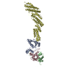
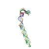
Mass: 62649.336 Da / Num. of mol.: 2 / Fragment: HEPHA4 ECTODOMAIN, RESIDUES 20-547
Source method: isolated from a genetically manipulated source
Source: (gene. exp.) HOMO SAPIENS (human) / Plasmid: PHLSEC / Cell line (production host): HEK293T / Production host: HOMO SAPIENS (human)
References: UniProt: P54764, receptor protein-tyrosine kinase
Sequence detailsSEQUENCE CORRESPONDS TO HEPHA4 RESIDUES 27-547 FUSED TO AN N-TERMINAL SECRETION SIGNAL SEQUENCE AND ...SEQUENCE CORRESPONDS TO HEPHA4 RESIDUES 27-547 FUSED TO AN N-TERMINAL SECRETION SIGNAL SEQUENCE AND A POLY-HISTIDINE TAG

-
Experimental details

-
Experiment

ExperimentMethod: X-RAY DIFFRACTION

-
Sample preparation

CrystalDensity Matthews: 6.16 Å3/Da / Density % sol: 76.7 %
Description: WE USED THE NEW CC CUTOFF CRITERIA FOR DETERMINING THE HIGH RES CUTOFF. CC-HALF IN THE HIGH RES SHELL IS 17.7
Crystal growDetails: 1.8 M AMMONIUM PHOSPHATE, 100 MM HEPES PH 7.4, ADDITIVE = EPHRINB2 IN A 1:1 MOLAR RATIO. WE SAW NO SIGN OF EPHRINB2 IN THE CRYSTAL STRUCTURE. THE USUAL BINDING SITE ON HEPHA4 WAS OCCUPIED BY ...Details: 1.8 M AMMONIUM PHOSPHATE, 100 MM HEPES PH 7.4, ADDITIVE = EPHRINB2 IN A 1:1 MOLAR RATIO. WE SAW NO SIGN OF EPHRINB2 IN THE CRYSTAL STRUCTURE. THE USUAL BINDING SITE ON HEPHA4 WAS OCCUPIED BY AN EPHA4-EPHA4 CRYSTAL CONTACT.

-
Data collection

Diffraction
IDMean temperature (K)Crystal-ID
11001
21001
Diffraction source
SourceSiteBeamlineIDWavelength
SYNCHROTRONDiamond I2410.9173
SYNCHROTRONDiamond I0420.9173
Radiation
IDProtocolMonochromatic (M) / Laue (L)Scattering typeWavelength-ID
1SINGLE WAVELENGTHMx-ray1
2SINGLE WAVELENGTHMx-ray1
Radiation wavelengthWavelength: 0.9173 Å / Relative weight: 1
ReflectionResolution: 3.65→83 Å / Num. obs: 34863 / % possible obs: 99.9 % / Observed criterion σ(I): 0 / Redundancy: 20 % / Biso Wilson estimate: 144.4 Å2 / Rmerge(I) obs: 0.19 / Net I/σ(I): 14.7
Reflection shellResolution: 3.65→3.7 Å / Redundancy: 20 % / Mean I/σ(I) obs: 0.48 / % possible all: 100

-
Processing

Software
NameVersionClassification
BUSTER2.11.2refinement
xia2data reduction
XDSdata reduction
xia2data scaling
XSCALEdata scaling
PHASERphasing
RefinementMethod to determine structure: MOLECULAR REPLACEMENT
Starting model: PDB ENTRY 2X10
Resolution: 3.65→48.18 Å / Cor.coef. Fo:Fc: 0.7358 / Cor.coef. Fo:Fc free: 0.6986 / SU R Cruickshank DPI: 1.041 / Cross valid method: THROUGHOUT / σ(F): 0 / SU R Blow DPI: 0.995 / SU Rfree Blow DPI: 0.584 / SU Rfree Cruickshank DPI: 0.599
RfactorNum. reflection% reflectionSelection details
Rfree0.3858 1753 5.03 %RANDOM
Rwork0.3495 ---
obs0.3513 34842 99.91 %-
Displacement parametersBiso mean: 124.97 Å2
Baniso -1Baniso -2Baniso -3
1--7.8639 Å20 Å20 Å2
2---7.8639 Å20 Å2
3---15.7278 Å2
Refine analyzeLuzzati coordinate error obs: 1.667 Å
Refinement stepCycle: LAST / Resolution: 3.65→48.18 Å
ProteinNucleic acidLigandSolventTotal
Num. atoms6344 0 0 0 6344
Refine LS restraints
Refine-IDTypeDev idealNumberRestraint functionWeight
X-RAY DIFFRACTIONt_bond_d0.0076496HARMONIC2
X-RAY DIFFRACTIONt_angle_deg0.858839HARMONIC2
X-RAY DIFFRACTIONt_dihedral_angle_d2230SINUSOIDAL2
X-RAY DIFFRACTIONt_incorr_chiral_ct
X-RAY DIFFRACTIONt_pseud_angle
X-RAY DIFFRACTIONt_trig_c_planes180HARMONIC2
X-RAY DIFFRACTIONt_gen_planes936HARMONIC5
X-RAY DIFFRACTIONt_it6496HARMONIC20
X-RAY DIFFRACTIONt_nbd
X-RAY DIFFRACTIONt_omega_torsion1.21
X-RAY DIFFRACTIONt_other_torsion21.35
X-RAY DIFFRACTIONt_improper_torsion
X-RAY DIFFRACTIONt_chiral_improper_torsion861SEMIHARMONIC5
X-RAY DIFFRACTIONt_sum_occupancies
X-RAY DIFFRACTIONt_utility_distance
X-RAY DIFFRACTIONt_utility_angle
X-RAY DIFFRACTIONt_utility_torsion
X-RAY DIFFRACTIONt_ideal_dist_contact6305SEMIHARMONIC4
LS refinement shellResolution: 3.65→3.76 Å / Total num. of bins used: 17
RfactorNum. reflection% reflection
Rfree0.2201 133 4.51 %
Rwork0.2057 2818 -
all0.2064 2951 -
obs--99.91 %
Refinement TLS params.

Method: refined / Refine-ID: X-RAY DIFFRACTION

IDL112)L122)L132)L222)L232)L332)S11 (Å °)S12 (Å °)S13 (Å °)S21 (Å °)S22 (Å °)S23 (Å °)S31 (Å °)S32 (Å °)S33 (Å °)T112)T122)T132)T222)T232)T332)Origin x (Å)Origin y (Å)Origin z (Å)
10.4879-0.3543-0.44930.05340.21510.20470.1445-0.08910.03740.0842-0.05940.0671-0.02620.0635-0.08510.58660.10240.06660.54780.03490.534-25.934659.929341.8729
2-0.25350.99190.18231.35280.21840.1742-0.0727-0.09890.32210.0802-0.0440.1660.22960.01350.11670.54170.0802-0.2070.5672-0.15390.6483-8.83711.2763-6.345
Refinement TLS group
IDRefine-IDRefine TLS-IDSelection details
1X-RAY DIFFRACTION1CHAIN A
2X-RAY DIFFRACTION2CHAIN B

+
About Yorodumi

-
News

-
Feb 9, 2022. New format data for meta-information of EMDB entries

New format data for meta-information of EMDB entries

  • Version 3 of the EMDB header file is now the official format.
  • The previous official version 1.9 will be removed from the archive.

Related info.:EMDB header

External links:wwPDB to switch to version 3 of the EMDB data model

-
Aug 12, 2020. Covid-19 info

Covid-19 info

URL: https://pdbj.org/emnavi/covid19.php

New page: Covid-19 featured information page in EM Navigator.

Related info.:Covid-19 info / Mar 5, 2020. Novel coronavirus structure data

+
Mar 5, 2020. Novel coronavirus structure data

Novel coronavirus structure data

Related info.:Yorodumi Speices / Aug 12, 2020. Covid-19 info

External links:COVID-19 featured content - PDBj / Molecule of the Month (242):Coronavirus Proteases

+
Jan 31, 2019. EMDB accession codes are about to change! (news from PDBe EMDB page)

EMDB accession codes are about to change! (news from PDBe EMDB page)

  • The allocation of 4 digits for EMDB accession codes will soon come to an end. Whilst these codes will remain in use, new EMDB accession codes will include an additional digit and will expand incrementally as the available range of codes is exhausted. The current 4-digit format prefixed with “EMD-” (i.e. EMD-XXXX) will advance to a 5-digit format (i.e. EMD-XXXXX), and so on. It is currently estimated that the 4-digit codes will be depleted around Spring 2019, at which point the 5-digit format will come into force.
  • The EM Navigator/Yorodumi systems omit the EMD- prefix.

Related info.:Q: What is EMD? / ID/Accession-code notation in Yorodumi/EM Navigator

External links:EMDB Accession Codes are Changing Soon! / Contact to PDBj

+
Jul 12, 2017. Major update of PDB

Major update of PDB

  • wwPDB released updated PDB data conforming to the new PDBx/mmCIF dictionary.
  • This is a major update changing the version number from 4 to 5, and with Remediation, in which all the entries are updated.
  • In this update, many items about electron microscopy experimental information are reorganized (e.g. em_software).
  • Now, EM Navigator and Yorodumi are based on the updated data.

External links:wwPDB Remediation / Enriched Model Files Conforming to OneDep Data Standards Now Available in the PDB FTP Archive

-
Yorodumi

Thousand views of thousand structures

  • Yorodumi is a browser for structure data from EMDB, PDB, SASBDB, etc.
  • This page is also the successor to EM Navigator detail page, and also detail information page/front-end page for Omokage search.
  • The word "yorodu" (or yorozu) is an old Japanese word meaning "ten thousand". "mi" (miru) is to see.

Related info.:EMDB / PDB / SASBDB / Comparison of 3 databanks / Yorodumi Search / Aug 31, 2016. New EM Navigator & Yorodumi / Yorodumi Papers / Jmol/JSmol / Function and homology information / Changes in new EM Navigator and Yorodumi

Read more