 Movie
Movie Controller

[English] 日本語
 Yorodumi
- PDB-4loh: Crystal structure of hSTING(H232) in complex with c[G(2',5')pA(3'... -
+ Open data

- Basic information
| Entry | Database: PDB / ID: 4loh | ||||||
|---|---|---|---|---|---|---|---|
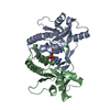
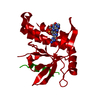
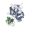
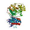
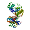
| Title | Crystal structure of hSTING(H232) in complex with c[G(2',5')pA(3',5')p] | ||||||
 Components Components | Stimulator of interferon genes protein | ||||||
 Keywords Keywords | IMMUNE SYSTEM / innate immunity | ||||||
| Function / homology |  Function and homology information Function and homology informationSTING complex / STAT6-mediated induction of chemokines / serine/threonine protein kinase complex / 2',3'-cyclic GMP-AMP binding / proton channel activity / cyclic-di-GMP binding / STING mediated induction of host immune responses / IRF3-mediated induction of type I IFN / cGAS/STING signaling pathway / positive regulation of type I interferon-mediated signaling pathway ...STING complex / STAT6-mediated induction of chemokines / serine/threonine protein kinase complex / 2',3'-cyclic GMP-AMP binding / proton channel activity / cyclic-di-GMP binding / STING mediated induction of host immune responses / IRF3-mediated induction of type I IFN / cGAS/STING signaling pathway / positive regulation of type I interferon-mediated signaling pathway / reticulophagy / pattern recognition receptor signaling pathway / cellular response to exogenous dsRNA / cytoplasmic pattern recognition receptor signaling pathway / autophagosome membrane / antiviral innate immune response / positive regulation of macroautophagy / autophagosome assembly / cellular response to organic cyclic compound / positive regulation of type I interferon production / autophagosome / cellular response to interferon-beta / signaling adaptor activity / positive regulation of defense response to virus by host / Regulation of innate immune responses to cytosolic DNA / endoplasmic reticulum-Golgi intermediate compartment membrane / activation of innate immune response / positive regulation of interferon-beta production / secretory granule membrane / cytoplasmic vesicle membrane / peroxisome / positive regulation of DNA-binding transcription factor activity / SARS-CoV-1 activates/modulates innate immune responses / positive regulation of protein binding / protein complex oligomerization / regulation of inflammatory response / defense response to virus / mitochondrial outer membrane / RNA polymerase II-specific DNA-binding transcription factor binding / endosome / Golgi membrane / innate immune response / ubiquitin protein ligase binding / Neutrophil degranulation / endoplasmic reticulum membrane / protein kinase binding / SARS-CoV-2 activates/modulates innate and adaptive immune responses / perinuclear region of cytoplasm / protein homodimerization activity / positive regulation of transcription by RNA polymerase II / nucleoplasm / identical protein binding / plasma membrane / cytosolSimilarity search - Function | ||||||
| Biological species |  Homo sapiens (human) Homo sapiens (human) | ||||||
| Method | X-RAY DIFFRACTION / SYNCHROTRON / MOLECULAR REPLACEMENT / Resolution: 2.25 Å | ||||||
 Authors Authors | Gao, P. / Patel, D.J. | ||||||
 Citation Citation | Journal: Cell(Cambridge,Mass.) / Year: 2013 Title: Structure-Function Analysis of STING Activation by c[G(2',5')pA(3',5')p] and Targeting by Antiviral DMXAA. Authors: Gao, P. / Ascano, M. / Zillinger, T. / Wang, W. / Dai, P. / Serganov, A.A. / Gaffney, B.L. / Shuman, S. / Jones, R.A. / Deng, L. / Hartmann, G. / Barchet, W. / Tuschl, T. / Patel, D.J. | ||||||
| History |
|
- Structure visualization
| Structure viewer | Molecule: MolmilJmol/JSmol |
|---|
- Downloads & links
-Download
| PDBx/mmCIF format | 4loh.cif.gz | 91.6 KB | Display | PDBx/mmCIF format |
|---|---|---|---|---|
| PDB format | pdb4loh.ent.gz | 70.5 KB | Display | PDB format |
| PDBx/mmJSON format | 4loh.json.gz | Tree view | PDBx/mmJSON format | |
| Others |  Other downloads Other downloads |
-Validation report
| Arichive directory | https://data.pdbj.org/pub/pdb/validation_reports/lo/4lohftp://data.pdbj.org/pub/pdb/validation_reports/lo/4loh | HTTPS FTP |
|---|
-Related structure data


-Links
 PDBj
PDBj


- Assembly
| Deposited unit | 
| ||||||||
|---|---|---|---|---|---|---|---|---|---|
| 1 |
| ||||||||
| Unit cell |
|
-Components
| #1: Protein | / hSTING / Endoplasmic reticulum interferon stimulator / ERIS / Mediator of IRF3 activation / hMITA / ...hSTING / Endoplasmic reticulum interferon stimulator / ERIS / Mediator of IRF3 activation / hMITA / Transmembrane protein 173 Mass: 21524.158 Da / Num. of mol.: 2 / Fragment: c-di-GMP-binding domain Source method: isolated from a genetically manipulated source Source: (gene. exp.) Homo sapiens (human) / Gene: TMEM173, ERIS, MITA, STING / Production host:  Escherichia coli (E. coli) / References: UniProt: Q86WV6 Escherichia coli (E. coli) / References: UniProt: Q86WV6#2: Chemical | ChemComp-1SY / | Cyclic guanosine monophosphate–adenosine monophosphate  Mass: 674.411 Da / Num. of mol.: 1 / Source method: obtained synthetically / Formula: C20H24N10O13P2 Mass: 674.411 Da / Num. of mol.: 1 / Source method: obtained synthetically / Formula: C20H24N10O13P2#3: Water | ChemComp-HOH / | Water Mass: 18.015 Da / Num. of mol.: 125 / Source method: isolated from a natural source / Formula: H2O Mass: 18.015 Da / Num. of mol.: 125 / Source method: isolated from a natural source / Formula: H2O |
|---|
-Experimental details
-Experiment
| Experiment | Method: X-RAY DIFFRACTION / Number of used crystals: 1 |
|---|
- Sample preparation
| Crystal | Density Matthews: 2.94 Å3/Da / Density % sol: 58.19 % |
|---|---|
| Crystal grow | Temperature: 293 K / Method: vapor diffusion, sitting drop / pH: 8.5 Details: 0.01 M NiCl2, 0.1 M Tris, 20% PEG2000, pH 8.5, VAPOR DIFFUSION, SITTING DROP, temperature 293K |
-Data collection
| Diffraction | Mean temperature: 200 K |
|---|---|
| Diffraction source | Source: SYNCHROTRON / Site: NSLS  / Beamline: X29A / Wavelength: 1.075 Å / Beamline: X29A / Wavelength: 1.075 Å |
| Detector | Type: ADSC QUANTUM 315r / Detector: CCD / Date: Apr 21, 2013 |
| Radiation | Monochromator: Si 111 CHANNEL / Protocol: SINGLE WAVELENGTH / Monochromatic (M) / Laue (L): M / Scattering type: x-ray |
| Radiation wavelength | Wavelength: 1.075 Å / Relative weight: 1 |
| Reflection | Resolution: 2.25→50 Å / Num. all: 21987 / Num. obs: 21448 / % possible obs: 97.55 % / Observed criterion σ(F): 2 / Observed criterion σ(I): 2 |
| Reflection shell | Resolution: 2.25→2.33 Å / % possible all: 97.1 |
- Processing
| Software |
| ||||||||||||||||||||||||||||||||||||||||||||||||||||||||||||||||||||||||||||||||||||||||||||||||||||||||||||||||||||||||||||||||||||||||||||||||||||||||||||||||||||||||||||||||||||||
|---|---|---|---|---|---|---|---|---|---|---|---|---|---|---|---|---|---|---|---|---|---|---|---|---|---|---|---|---|---|---|---|---|---|---|---|---|---|---|---|---|---|---|---|---|---|---|---|---|---|---|---|---|---|---|---|---|---|---|---|---|---|---|---|---|---|---|---|---|---|---|---|---|---|---|---|---|---|---|---|---|---|---|---|---|---|---|---|---|---|---|---|---|---|---|---|---|---|---|---|---|---|---|---|---|---|---|---|---|---|---|---|---|---|---|---|---|---|---|---|---|---|---|---|---|---|---|---|---|---|---|---|---|---|---|---|---|---|---|---|---|---|---|---|---|---|---|---|---|---|---|---|---|---|---|---|---|---|---|---|---|---|---|---|---|---|---|---|---|---|---|---|---|---|---|---|---|---|---|---|---|---|---|---|
| Refinement | Method to determine structure: MOLECULAR REPLACEMENT / Resolution: 2.25→31.87 Å / Cor.coef. Fo:Fc: 0.943 / Cor.coef. Fo:Fc free: 0.928 / SU B: 4.915 / SU ML: 0.123 / Cross valid method: THROUGHOUT / σ(F): 2 / ESU R: 0.26 / ESU R Free: 0.192 / Stereochemistry target values: MAXIMUM LIKELIHOOD / Details: HYDROGENS HAVE BEEN ADDED IN THE RIDING POSITIONS
| ||||||||||||||||||||||||||||||||||||||||||||||||||||||||||||||||||||||||||||||||||||||||||||||||||||||||||||||||||||||||||||||||||||||||||||||||||||||||||||||||||||||||||||||||||||||
| Solvent computation | Ion probe radii: 0.8 Å / Shrinkage radii: 0.8 Å / VDW probe radii: 1.2 Å / Solvent model: MASK | ||||||||||||||||||||||||||||||||||||||||||||||||||||||||||||||||||||||||||||||||||||||||||||||||||||||||||||||||||||||||||||||||||||||||||||||||||||||||||||||||||||||||||||||||||||||
| Displacement parameters | Biso mean: 34.78 Å2
| ||||||||||||||||||||||||||||||||||||||||||||||||||||||||||||||||||||||||||||||||||||||||||||||||||||||||||||||||||||||||||||||||||||||||||||||||||||||||||||||||||||||||||||||||||||||
| Refinement step | Cycle: LAST / Resolution: 2.25→31.87 Å
| ||||||||||||||||||||||||||||||||||||||||||||||||||||||||||||||||||||||||||||||||||||||||||||||||||||||||||||||||||||||||||||||||||||||||||||||||||||||||||||||||||||||||||||||||||||||
| Refine LS restraints |
|