 Movie
Movie Controller


+ Open data

- Basic information
| Entry | Database: PDB / ID: 4xoy | |||||||||
|---|---|---|---|---|---|---|---|---|---|---|
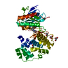
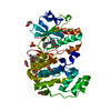
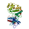
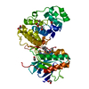
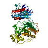
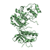
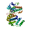
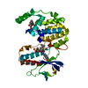
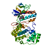
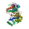
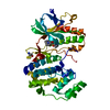
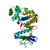
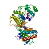
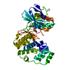
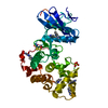
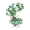
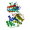
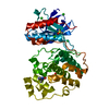
| Title | Crystal structure of ERK2 in complex with an inhibitor | |||||||||
 Components Components | Mitogen-activated protein kinase 1 | |||||||||
 Keywords Keywords |  TRANSFERASE / SERINE/THREONINE-PROTEIN KINASE TRANSFERASE / SERINE/THREONINE-PROTEIN KINASE | |||||||||
| Function / homology |  Function and homology information Function and homology informationphospho-PLA2 pathway / RAF-independent MAPK1/3 activation / MAPK1 (ERK2) activation / Signaling by NODAL / Frs2-mediated activation / ERK/MAPK targets / ERKs are inactivated / Activation of the AP-1 family of transcription factors / RHO GTPases Activate WASPs and WAVEs / IFNG signaling activates MAPKs ...phospho-PLA2 pathway / RAF-independent MAPK1/3 activation / MAPK1 (ERK2) activation / Signaling by NODAL / Frs2-mediated activation / ERK/MAPK targets / ERKs are inactivated / Activation of the AP-1 family of transcription factors / RHO GTPases Activate WASPs and WAVEs / IFNG signaling activates MAPKs / Negative feedback regulation of MAPK pathway / Gastrin-CREB signalling pathway via PKC and MAPK / Estrogen-dependent nuclear events downstream of ESR-membrane signaling / Golgi Cisternae Pericentriolar Stack Reorganization / Regulation of actin dynamics for phagocytic cup formation / Estrogen-stimulated signaling through PRKCZ / Growth hormone receptor signaling / Spry regulation of FGF signaling / SMAD2/SMAD3:SMAD4 heterotrimer regulates transcription / Oxidative Stress Induced Senescence / Senescence-Associated Secretory Phenotype (SASP) / Oncogene Induced Senescence / Signaling by Activin / Downregulation of SMAD2/3:SMAD4 transcriptional activity / Signal attenuation / NCAM signaling for neurite out-growth / Negative regulation of FGFR1 signaling / Negative regulation of FGFR3 signaling / Negative regulation of FGFR4 signaling / Regulation of the apoptosome activity / Signal transduction by L1 / Negative regulation of FGFR2 signaling / RHO GTPases Activate NADPH Oxidases / Negative regulation of MAPK pathway / Interferon gamma signaling / FCERI mediated MAPK activation / PI5P, PP2A and IER3 Regulate PI3K/AKT Signaling / Regulation of HSF1-mediated heat shock response / MAP2K and MAPK activation / diadenosine tetraphosphate biosynthetic process / Recycling pathway of L1 / neural crest cell development / cardiac neural crest cell development involved in heart development / caveolin-mediated endocytosis / cytosine metabolic process / response to epidermal growth factor / mitogen-activated protein kinase kinase kinase binding / regulation of cellular pH / positive regulation of macrophage proliferation / outer ear morphogenesis / Thrombin signalling through proteinase activated receptors (PARs) / RAF/MAP kinase cascade / regulation of Golgi inheritance / ERBB signaling pathway / labyrinthine layer blood vessel development / Neutrophil degranulation / mammary gland epithelial cell proliferation / trachea formation / regulation of early endosome to late endosome transport / : / regulation of stress-activated MAPK cascade / positive regulation of macrophage chemotaxis / lung morphogenesis / ERBB2-ERBB3 signaling pathway / response to exogenous dsRNA / regulation of cytoskeleton organization / face development / androgen receptor signaling pathway / pseudopodium / progesterone receptor signaling pathway / negative regulation of cell differentiation / Bergmann glial cell differentiation / positive regulation of telomere capping / thyroid gland development / decidualization / steroid hormone mediated signaling pathway / MAP kinase activity / regulation of ossification / mitogen-activated protein kinase / phosphatase binding / Schwann cell development / stress-activated MAPK cascade / lipopolysaccharide-mediated signaling pathway / positive regulation of cardiac muscle cell proliferation / sensory perception of pain / cellular response to cadmium ion / positive regulation of telomere maintenance via telomerase / ERK1 and ERK2 cascade / cellular response to amino acid starvation / myelination / dendrite cytoplasm / phosphotyrosine residue binding / RNA polymerase II CTD heptapeptide repeat kinase activity / insulin-like growth factor receptor signaling pathway / thymus development / positive regulation of peptidyl-threonine phosphorylation / caveola / positive regulation of translation / long-term synaptic potentiation / animal organ morphogenesisSimilarity search - Function | |||||||||
| Biological species |  Rattus norvegicus (Norway rat) Rattus norvegicus (Norway rat) | |||||||||
| Method | X-RAY DIFFRACTION / SYNCHROTRON / MIR / Resolution: 2.1 Å | |||||||||
 Authors Authors | Gelin, M. / Allemand, F. / Labesse, G. / Guichou, J.F. | |||||||||
| Funding support |  France, 1items France, 1items
| |||||||||
 Citation Citation | Journal: Acta Crystallogr.,Sect.D / Year: 2015 Title: Combining `dry' co-crystallization and in situ diffraction to facilitate ligand screening by X-ray crystallography. Authors: Gelin, M. / Delfosse, V. / Allemand, F. / Hoh, F. / Sallaz-Damaz, Y. / Pirocchi, M. / Bourguet, W. / Ferrer, J.L. / Labesse, G. / Guichou, J.F. | |||||||||
| History |
|
- Structure visualization
| Structure viewer | Molecule: MolmilJmol/JSmol |
|---|
- Downloads & links
-Download
| PDBx/mmCIF format | 4xoy.cif.gz | 149.7 KB | Display | PDBx/mmCIF format |
|---|---|---|---|---|
| PDB format | pdb4xoy.ent.gz | 116.1 KB | Display | PDB format |
| PDBx/mmJSON format | 4xoy.json.gz | Tree view | PDBx/mmJSON format | |
| Others |  Other downloads Other downloads |
-Validation report
| Arichive directory | https://data.pdbj.org/pub/pdb/validation_reports/xo/4xoyftp://data.pdbj.org/pub/pdb/validation_reports/xo/4xoy | HTTPS FTP |
|---|
-Related structure data
| Related structure data |  3rdcC 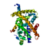 4xldC  4xn6C  4xncC  4xneC  4xozC  4xp0C  4xp2C  4xp3C  4xrjC  4xrlC  4zscC 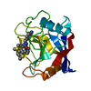 4zsdC  3qywS S: Starting model for refinement C: citing same article ( |
|---|---|
| Similar structure data |


-Links
 PDBj
PDBj


- Assembly
| Deposited unit | 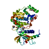
| ||||||||
|---|---|---|---|---|---|---|---|---|---|
| 1 |
| ||||||||
| Unit cell |
| ||||||||
| Details | biological unit is the same as asym. |
-Components
| #1: Protein | Mass: 41077.367 Da / Num. of mol.: 1 / Fragment: UNP residues 8-358 Source method: isolated from a genetically manipulated source Source: (gene. exp.) Rattus norvegicus (Norway rat) / Gene: Mapk1, Erk2, Mapk, Prkm1 / Production host:  Escherichia coli (E. coli) Escherichia coli (E. coli)References: UniProt: P63086, mitogen-activated protein kinase |
|---|---|
| #2: Chemical | ChemComp-DX4 / Tioguanine  Mass: 167.192 Da / Num. of mol.: 1 Mass: 167.192 Da / Num. of mol.: 1Source method: isolated from a genetically manipulated source Formula: C5H5N5S / Comment: medication*YM |
| #3: Chemical | ChemComp-SO4 / Sulfate  Mass: 96.063 Da / Num. of mol.: 1 / Source method: isolated from a natural source / Formula: SO4 Mass: 96.063 Da / Num. of mol.: 1 / Source method: isolated from a natural source / Formula: SO4 |
| #4: Water | ChemComp-HOH / Water Mass: 18.015 Da / Num. of mol.: 143 / Source method: isolated from a natural source / Formula: H2O Mass: 18.015 Da / Num. of mol.: 143 / Source method: isolated from a natural source / Formula: H2O |
-Experimental details
-Experiment
| Experiment | Method: X-RAY DIFFRACTION / Number of used crystals: 1 |
|---|
- Sample preparation
| Crystal | Density Matthews: 2.48 Å3/Da / Density % sol: 50.4 % |
|---|---|
| Crystal grow | Temperature: 291 K / Method: vapor diffusion, hanging drop / pH: 6.5 Details: 26% PEG MME 2000, 0.1M MES pH 6.5, 0.1M ammonium sulfate, 0.02M beta-mercaptoethanol |
-Data collection
| Diffraction | Mean temperature: 298 K |
|---|---|
| Diffraction source | Source: SYNCHROTRON / Site: ESRF / Beamline: BM30A / Wavelength: 0.97922 Å |
| Detector | Type: ADSC QUANTUM 315r / Detector: CCD / Date: Jul 21, 2014 |
| Radiation | Protocol: SINGLE WAVELENGTH / Monochromatic (M) / Laue (L): M / Scattering type: x-ray |
| Radiation wavelength | Wavelength: 0.97922 Å / Relative weight: 1 |
| Reflection | Resolution: 2.1→29.305 Å / Num. obs: 28103 / % possible obs: 89.8 % / Redundancy: 2.5 % / Net I/σ(I): 6 |
| Reflection shell | Resolution: 2.1→2.15 Å / Rmerge(I) obs: 0.514 / Mean I/σ(I) obs: 1.95 / % possible all: 67.8 |
- Processing
| Software |
| |||||||||||||||||||||||||||||||||||||||||||||||||||||||||||||||||||||||||||||||||||||||||||||||||||||||||||||||||||||||||||||||||||||||||||||||||||||||||||||||||||||||||||||||
|---|---|---|---|---|---|---|---|---|---|---|---|---|---|---|---|---|---|---|---|---|---|---|---|---|---|---|---|---|---|---|---|---|---|---|---|---|---|---|---|---|---|---|---|---|---|---|---|---|---|---|---|---|---|---|---|---|---|---|---|---|---|---|---|---|---|---|---|---|---|---|---|---|---|---|---|---|---|---|---|---|---|---|---|---|---|---|---|---|---|---|---|---|---|---|---|---|---|---|---|---|---|---|---|---|---|---|---|---|---|---|---|---|---|---|---|---|---|---|---|---|---|---|---|---|---|---|---|---|---|---|---|---|---|---|---|---|---|---|---|---|---|---|---|---|---|---|---|---|---|---|---|---|---|---|---|---|---|---|---|---|---|---|---|---|---|---|---|---|---|---|---|---|---|---|---|---|
| Refinement | Method to determine structure: MIR Starting model: Isomorphous replacement with a simple Rigid Body with Refmac5, using another structure of the same proteine (3QYW) Resolution: 2.1→29.287 Å / SU ML: 0.24 / Cross valid method: FREE R-VALUE / σ(F): 0.88 / Phase error: 22.67 / Stereochemistry target values: ML
| |||||||||||||||||||||||||||||||||||||||||||||||||||||||||||||||||||||||||||||||||||||||||||||||||||||||||||||||||||||||||||||||||||||||||||||||||||||||||||||||||||||||||||||||
| Solvent computation | Shrinkage radii: 0.9 Å / VDW probe radii: 1.11 Å / Solvent model: FLAT BULK SOLVENT MODEL | |||||||||||||||||||||||||||||||||||||||||||||||||||||||||||||||||||||||||||||||||||||||||||||||||||||||||||||||||||||||||||||||||||||||||||||||||||||||||||||||||||||||||||||||
| Refinement step | Cycle: LAST / Resolution: 2.1→29.287 Å
| |||||||||||||||||||||||||||||||||||||||||||||||||||||||||||||||||||||||||||||||||||||||||||||||||||||||||||||||||||||||||||||||||||||||||||||||||||||||||||||||||||||||||||||||
| Refine LS restraints |
| |||||||||||||||||||||||||||||||||||||||||||||||||||||||||||||||||||||||||||||||||||||||||||||||||||||||||||||||||||||||||||||||||||||||||||||||||||||||||||||||||||||||||||||||
| LS refinement shell |
| |||||||||||||||||||||||||||||||||||||||||||||||||||||||||||||||||||||||||||||||||||||||||||||||||||||||||||||||||||||||||||||||||||||||||||||||||||||||||||||||||||||||||||||||
| Refinement TLS params. | Method: refined / Refine-ID: X-RAY DIFFRACTION
| |||||||||||||||||||||||||||||||||||||||||||||||||||||||||||||||||||||||||||||||||||||||||||||||||||||||||||||||||||||||||||||||||||||||||||||||||||||||||||||||||||||||||||||||
| Refinement TLS group |
|
