 Movie
Movie Controller

[English] 日本語
 Yorodumi
- PDB-4lh2: Structure of mouse 1-Pyrroline-5-Carboxylate Dehydrogenase (ALDH4... -
+ Open data

- Basic information
| Entry | Database: PDB / ID: 4lh2 | ||||||
|---|---|---|---|---|---|---|---|
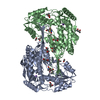
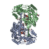
| Title | Structure of mouse 1-Pyrroline-5-Carboxylate Dehydrogenase (ALDH4A1) complexed with succinate | ||||||
 Components Components | Delta-1-pyrroline-5-carboxylate dehydrogenase, mitochondrial | ||||||
 Keywords Keywords |  OXIDOREDUCTASE / Proline catabolism / Substrate recognition / Rossmann Fold OXIDOREDUCTASE / Proline catabolism / Substrate recognition / Rossmann Fold | ||||||
| Function / homology |  Function and homology information Function and homology informationProline catabolism / Glyoxylate metabolism and glycine degradation / 1-pyrroline-5-carboxylate dehydrogenase activity / L-glutamate gamma-semialdehyde dehydrogenase / proline catabolic process to glutamate / aldehyde dehydrogenase (NAD+) activity / mitochondrial matrix / mitochondrion / identical protein binding / cytosolSimilarity search - Function | ||||||
| Biological species |  Mus musculus (house mouse) Mus musculus (house mouse) | ||||||
| Method | X-RAY DIFFRACTION / FOURIER SYNTHESIS / Resolution: 1.673 Å | ||||||
 Authors Authors | Pemberton, T.A. / Tanner, J.J. | ||||||
 Citation Citation | Journal: Arch.Biochem.Biophys. / Year: 2013 Title: Structural basis of substrate selectivity of Delta (1)-pyrroline-5-carboxylate dehydrogenase (ALDH4A1): Semialdehyde chain length. Authors: Pemberton, T.A. / Tanner, J.J. | ||||||
| History |
|
- Structure visualization
| Structure viewer | Molecule: MolmilJmol/JSmol |
|---|
- Downloads & links
-Download
| PDBx/mmCIF format | 4lh2.cif.gz | 417.5 KB | Display | PDBx/mmCIF format |
|---|---|---|---|---|
| PDB format | pdb4lh2.ent.gz | 336.6 KB | Display | PDB format |
| PDBx/mmJSON format | 4lh2.json.gz | Tree view | PDBx/mmJSON format | |
| Others |  Other downloads Other downloads |
-Validation report
| Arichive directory | https://data.pdbj.org/pub/pdb/validation_reports/lh/4lh2ftp://data.pdbj.org/pub/pdb/validation_reports/lh/4lh2 | HTTPS FTP |
|---|
-Related structure data
| Related structure data | 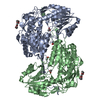 4lgzC 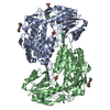 4lh0C 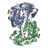 4lh1C  4lh3C 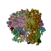 4v9jS C: citing same article ( S: Starting model for refinement |
|---|---|
| Similar structure data |


-Links
 PDBj
PDBj

- Assembly
| Deposited unit | 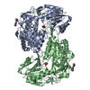
| ||||||||
|---|---|---|---|---|---|---|---|---|---|
| 1 |
| ||||||||
| Unit cell |
|
-Components
| #1: Protein | Mass: 61954.215 Da / Num. of mol.: 2 / Fragment: unp residues 21-562 Source method: isolated from a genetically manipulated source Source: (gene. exp.) Mus musculus (house mouse) / Gene: ADLH4a1, Aldh4a1 / Plasmid: pET28a / Production host:  Escherichia coli (E. coli) / References: UniProt: Q8CHT0, EC: 1.5.1.12 Escherichia coli (E. coli) / References: UniProt: Q8CHT0, EC: 1.5.1.12#2: Chemical | Polyethylene glycol  Mass: 238.278 Da / Num. of mol.: 2 / Source method: obtained synthetically / Formula: C10H22O6 / Comment: precipitant*YM Mass: 238.278 Da / Num. of mol.: 2 / Source method: obtained synthetically / Formula: C10H22O6 / Comment: precipitant*YM#3: Chemical | Succinic acid  Mass: 118.088 Da / Num. of mol.: 2 / Source method: obtained synthetically / Formula: C4H6O4 Mass: 118.088 Da / Num. of mol.: 2 / Source method: obtained synthetically / Formula: C4H6O4#4: Water | ChemComp-HOH / | Water Mass: 18.015 Da / Num. of mol.: 799 / Source method: isolated from a natural source / Formula: H2O Mass: 18.015 Da / Num. of mol.: 799 / Source method: isolated from a natural source / Formula: H2O |
|---|
-Experimental details
-Experiment
| Experiment | Method: X-RAY DIFFRACTION / Number of used crystals: 1 |
|---|
- Sample preparation
| Crystal | Density Matthews: 2.13 Å3/Da / Density % sol: 42.32 % |
|---|---|
| Crystal grow | Temperature: 298 K / Method: vapor diffusion, sitting drop / pH: 6.5 Details: 15-25% PEG 3350, 0.2 M lithium sulfate, 0.1 M Bis-Tris, pH 6.5, VAPOR DIFFUSION, SITTING DROP, temperature 298K |
-Data collection
| Diffraction | Mean temperature: 148 K | ||||||||||||||||||||||||||||||||||||||||||||||||||||||||||||||||||||||||||||||||||||||||
|---|---|---|---|---|---|---|---|---|---|---|---|---|---|---|---|---|---|---|---|---|---|---|---|---|---|---|---|---|---|---|---|---|---|---|---|---|---|---|---|---|---|---|---|---|---|---|---|---|---|---|---|---|---|---|---|---|---|---|---|---|---|---|---|---|---|---|---|---|---|---|---|---|---|---|---|---|---|---|---|---|---|---|---|---|---|---|---|---|---|
| Diffraction source | Source: ROTATING ANODE / Type: RIGAKU / Wavelength: 1.5418 Å | ||||||||||||||||||||||||||||||||||||||||||||||||||||||||||||||||||||||||||||||||||||||||
| Detector | Type: RIGAKU RAXIS IV++ / Detector: IMAGE PLATE / Date: Aug 24, 2012 | ||||||||||||||||||||||||||||||||||||||||||||||||||||||||||||||||||||||||||||||||||||||||
| Radiation | Monochromator: osmic blue / Protocol: SINGLE WAVELENGTH / Monochromatic (M) / Laue (L): M / Scattering type: x-ray | ||||||||||||||||||||||||||||||||||||||||||||||||||||||||||||||||||||||||||||||||||||||||
| Radiation wavelength | Wavelength: 1.5418 Å / Relative weight: 1 | ||||||||||||||||||||||||||||||||||||||||||||||||||||||||||||||||||||||||||||||||||||||||
| Reflection | Resolution: 1.673→19.677 Å / Num. all: 118798 / Num. obs: 118798 / % possible obs: 97 % / Redundancy: 4.6 % / Rsym value: 0.055 / Net I/σ(I): 16.3 | ||||||||||||||||||||||||||||||||||||||||||||||||||||||||||||||||||||||||||||||||||||||||
| Reflection shell | Diffraction-ID: 1
|
- Processing
| Software |
| |||||||||||||||||||||||||||||||||||||||||||||||||||||||||||||||||||||||||||||||||||||||||||||||||||||||||||||||||||||||||||||||||||||||||||||||||||||||||||||||||||||||||||||||||||||||||||||||||||||||||||||||||||||||||
|---|---|---|---|---|---|---|---|---|---|---|---|---|---|---|---|---|---|---|---|---|---|---|---|---|---|---|---|---|---|---|---|---|---|---|---|---|---|---|---|---|---|---|---|---|---|---|---|---|---|---|---|---|---|---|---|---|---|---|---|---|---|---|---|---|---|---|---|---|---|---|---|---|---|---|---|---|---|---|---|---|---|---|---|---|---|---|---|---|---|---|---|---|---|---|---|---|---|---|---|---|---|---|---|---|---|---|---|---|---|---|---|---|---|---|---|---|---|---|---|---|---|---|---|---|---|---|---|---|---|---|---|---|---|---|---|---|---|---|---|---|---|---|---|---|---|---|---|---|---|---|---|---|---|---|---|---|---|---|---|---|---|---|---|---|---|---|---|---|---|---|---|---|---|---|---|---|---|---|---|---|---|---|---|---|---|---|---|---|---|---|---|---|---|---|---|---|---|---|---|---|---|---|---|---|---|---|---|---|---|---|---|---|---|---|---|---|---|---|
| Refinement | Method to determine structure: FOURIER SYNTHESIS Starting model: pdb entry 4V9J Resolution: 1.673→19.677 Å / Occupancy max: 1 / Occupancy min: 0.29 / FOM work R set: 0.8904 / SU ML: 0.18 / Cross valid method: THROUGHOUT / σ(F): 1.04 / Phase error: 18.37 / Stereochemistry target values: ML
| |||||||||||||||||||||||||||||||||||||||||||||||||||||||||||||||||||||||||||||||||||||||||||||||||||||||||||||||||||||||||||||||||||||||||||||||||||||||||||||||||||||||||||||||||||||||||||||||||||||||||||||||||||||||||
| Solvent computation | Shrinkage radii: 0.83 Å / VDW probe radii: 1.1 Å / Solvent model: FLAT BULK SOLVENT MODEL / Bsol: 43.816 Å2 / ksol: 0.408 e/Å3 | |||||||||||||||||||||||||||||||||||||||||||||||||||||||||||||||||||||||||||||||||||||||||||||||||||||||||||||||||||||||||||||||||||||||||||||||||||||||||||||||||||||||||||||||||||||||||||||||||||||||||||||||||||||||||
| Displacement parameters | Biso max: 61.46 Å2 / Biso mean: 15.9034 Å2 / Biso min: 4.11 Å2
| |||||||||||||||||||||||||||||||||||||||||||||||||||||||||||||||||||||||||||||||||||||||||||||||||||||||||||||||||||||||||||||||||||||||||||||||||||||||||||||||||||||||||||||||||||||||||||||||||||||||||||||||||||||||||
| Refinement step | Cycle: LAST / Resolution: 1.673→19.677 Å
| |||||||||||||||||||||||||||||||||||||||||||||||||||||||||||||||||||||||||||||||||||||||||||||||||||||||||||||||||||||||||||||||||||||||||||||||||||||||||||||||||||||||||||||||||||||||||||||||||||||||||||||||||||||||||
| Refine LS restraints |
| |||||||||||||||||||||||||||||||||||||||||||||||||||||||||||||||||||||||||||||||||||||||||||||||||||||||||||||||||||||||||||||||||||||||||||||||||||||||||||||||||||||||||||||||||||||||||||||||||||||||||||||||||||||||||
| LS refinement shell | Refine-ID: X-RAY DIFFRACTION / Total num. of bins used: 30
| |||||||||||||||||||||||||||||||||||||||||||||||||||||||||||||||||||||||||||||||||||||||||||||||||||||||||||||||||||||||||||||||||||||||||||||||||||||||||||||||||||||||||||||||||||||||||||||||||||||||||||||||||||||||||
| Refinement TLS params. | Method: refined / Refine-ID: X-RAY DIFFRACTION
| |||||||||||||||||||||||||||||||||||||||||||||||||||||||||||||||||||||||||||||||||||||||||||||||||||||||||||||||||||||||||||||||||||||||||||||||||||||||||||||||||||||||||||||||||||||||||||||||||||||||||||||||||||||||||
| Refinement TLS group |
|
