 Movie
Movie Controller

[English] 日本語
 Yorodumi
- PDB-4ldn: Crystal structure of a putative purine nucleoside phosphorylase f... -
+ Open data

- Basic information
| Entry | Database: PDB / ID: 4ldn | ||||||
|---|---|---|---|---|---|---|---|
| Title | Crystal structure of a putative purine nucleoside phosphorylase from Vibrio fischeri ES114 (Target NYSGRC-029521) | ||||||
 Components Components | Purine nucleoside phosphorylase DeoD-type | ||||||
 Keywords Keywords |  TRANSFERASE / Structural genomics / NYSGRC / PSI-Biology / New York Structural Genomics Research Consortium TRANSFERASE / Structural genomics / NYSGRC / PSI-Biology / New York Structural Genomics Research Consortium | ||||||
| Function / homology |  Function and homology information Function and homology informationpurine nucleoside metabolic process / purine-nucleoside phosphorylase / purine-nucleoside phosphorylase activitySimilarity search - Function | ||||||
| Biological species |  Vibrio fischeri (bacteria) Vibrio fischeri (bacteria) | ||||||
| Method | X-RAY DIFFRACTION / SYNCHROTRON / SAD / Resolution: 1.48 Å | ||||||
 Authors Authors | Sampathkumar, P. / Almo, S.C. / New York Structural Genomics Research Consortium (NYSGRC) | ||||||
 Citation Citation | Journal: to be published Title: Crystal structure of a putative purine nucleoside phosphorylase from Vibrio fischeri ES114 (Target NYSGRC-029521) Authors: Sampathkumar, P. / Ahmed, M. / Attonito, J. / Bhosle, R. / Bonanno, J. / Chamala, S. / Chowdhury, S. / Eromenok, G. / Fiser, A. / Glenn, A.S. / Hammonds, J. / Himmel, D.M. / Hillerich, B. / ...Authors: Sampathkumar, P. / Ahmed, M. / Attonito, J. / Bhosle, R. / Bonanno, J. / Chamala, S. / Chowdhury, S. / Eromenok, G. / Fiser, A. / Glenn, A.S. / Hammonds, J. / Himmel, D.M. / Hillerich, B. / Khafizov, K. / Lafleur, J. / Love, J.D. / Stead, M. / Seidel, R. / Toro, R. / Morisco, L.L. / Sojitra, S.S. / Wasserman, S.R. / Suarez, J. / Schramm, V.L. / Almo, S.C. | ||||||
| History |
|
- Structure visualization
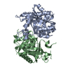
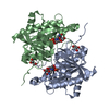
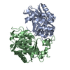
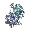
| Structure viewer | Molecule: MolmilJmol/JSmol |
|---|
- Downloads & links
-Download
| PDBx/mmCIF format | 4ldn.cif.gz | 63.8 KB | Display | PDBx/mmCIF format |
|---|---|---|---|---|
| PDB format | pdb4ldn.ent.gz | 51.6 KB | Display | PDB format |
| PDBx/mmJSON format | 4ldn.json.gz | Tree view | PDBx/mmJSON format | |
| Others |  Other downloads Other downloads |
-Validation report
| Arichive directory | https://data.pdbj.org/pub/pdb/validation_reports/ld/4ldnftp://data.pdbj.org/pub/pdb/validation_reports/ld/4ldn | HTTPS FTP |
|---|
-Related structure data
| Similar structure data | |
|---|---|
| Other databases |
-Links
 PDBj
PDBj- Assembly
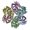
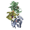
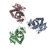
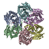
| Deposited unit | 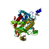
| ||||||||||||||||||
|---|---|---|---|---|---|---|---|---|---|---|---|---|---|---|---|---|---|---|---|
| 1 | x 6
| ||||||||||||||||||
| Unit cell |
| ||||||||||||||||||
| Components on special symmetry positions |
| ||||||||||||||||||
| Details | probable hexamer |
-Components
| #1: Protein | Mass: 29741.729 Da / Num. of mol.: 1 Source method: isolated from a genetically manipulated source Source: (gene. exp.) Vibrio fischeri (bacteria) / Strain: ES114 / Gene: deoD3, VF_A0968 / Plasmid: pSGC-His / Production host: Escherichia coli (E. coli) / Strain (production host): BL21(DE3) CodonPlus RILReferences: UniProt: Q5DYV8, purine-nucleoside phosphorylase | ||||
|---|---|---|---|---|---|
| #2: Chemical | Phosphate  Mass: 94.971 Da / Num. of mol.: 2 / Source method: obtained synthetically / Formula: PO4 Mass: 94.971 Da / Num. of mol.: 2 / Source method: obtained synthetically / Formula: PO4#3: Chemical | ChemComp-EDO / Ethylene glycol  Mass: 62.068 Da / Num. of mol.: 5 / Source method: obtained synthetically / Formula: C2H6O2 Mass: 62.068 Da / Num. of mol.: 5 / Source method: obtained synthetically / Formula: C2H6O2#4: Water | ChemComp-HOH / | Water Mass: 18.015 Da / Num. of mol.: 164 / Source method: isolated from a natural source / Formula: H2O Mass: 18.015 Da / Num. of mol.: 164 / Source method: isolated from a natural source / Formula: H2O |
-Experimental details
-Experiment
| Experiment | Method: X-RAY DIFFRACTION / Number of used crystals: 1 |
|---|
- Sample preparation
| Crystal | Density Matthews: 1.95 Å3/Da / Density % sol: 36.97 % |
|---|---|
| Crystal grow | Temperature: 298 K / Method: vapor diffusion, sitting drop / pH: 10.5 Details: Protein (20mM HEPES pH7.5, 150mM NaCl, 5% glycerol, and 5mM DTT), Reservoir (MCSG2 #05 - 0.2 M Lithium Sulfate, 0.1 M CAPS:NaOH pH 10.5, 1.2 M NaH2PO4/0.8 M K2HPO4), Cryoprotection (33% ...Details: Protein (20mM HEPES pH7.5, 150mM NaCl, 5% glycerol, and 5mM DTT), Reservoir (MCSG2 #05 - 0.2 M Lithium Sulfate, 0.1 M CAPS:NaOH pH 10.5, 1.2 M NaH2PO4/0.8 M K2HPO4), Cryoprotection (33% Ethylene glycol), VAPOR DIFFUSION, SITTING DROP, temperature 298K |
-Data collection
| Diffraction | Mean temperature: 100 K |
|---|---|
| Diffraction source | Source: SYNCHROTRON / Site: APS  / Beamline: 31-ID / Wavelength: 0.97931 Å / Beamline: 31-ID / Wavelength: 0.97931 Å |
| Detector | Type: RAYONIX MX225HE / Detector: CCD / Date: Apr 18, 2013 / Details: MIRRORS |
| Radiation | Monochromator: Diamond(111) / Protocol: SINGLE WAVELENGTH / Monochromatic (M) / Laue (L): M / Scattering type: x-ray |
| Radiation wavelength | Wavelength: 0.97931 Å / Relative weight: 1 |
| Reflection | Resolution: 1.48→40 Å / Num. obs: 37977 / % possible obs: 99.7 % / Observed criterion σ(I): 0 / Redundancy: 14.8 % / Biso Wilson estimate: 12 Å2 / Rmerge(I) obs: 0.112 / Net I/σ(I): 23.7 |
| Reflection shell | Resolution: 1.48→1.51 Å / Redundancy: 13.5 % / Rmerge(I) obs: 0.801 / Mean I/σ(I) obs: 3.7 / Num. unique all: 1837 / % possible all: 96.8 |
- Processing
| Software |
| |||||||||||||||||||||||||||||||||||||||||||||||||||||||||||||||||||||||||||
|---|---|---|---|---|---|---|---|---|---|---|---|---|---|---|---|---|---|---|---|---|---|---|---|---|---|---|---|---|---|---|---|---|---|---|---|---|---|---|---|---|---|---|---|---|---|---|---|---|---|---|---|---|---|---|---|---|---|---|---|---|---|---|---|---|---|---|---|---|---|---|---|---|---|---|---|---|
| Refinement | Method to determine structure: SAD / Resolution: 1.48→30.84 Å / Cor.coef. Fo:Fc: 0.972 / Cor.coef. Fo:Fc free: 0.964 / Occupancy max: 1 / Occupancy min: 0.5 / SU B: 1.41 / SU ML: 0.052 / Cross valid method: THROUGHOUT / σ(F): 0 / ESU R: 0.069 / ESU R Free: 0.069 / Stereochemistry target values: MAXIMUM LIKELIHOOD Details: HYDROGENS HAVE BEEN ADDED IN THE RIDING POSITIONS U VALUES : REFINED INDIVIDUALLY
| |||||||||||||||||||||||||||||||||||||||||||||||||||||||||||||||||||||||||||
| Solvent computation | Ion probe radii: 0.8 Å / Shrinkage radii: 0.8 Å / VDW probe radii: 1.4 Å / Solvent model: MASK | |||||||||||||||||||||||||||||||||||||||||||||||||||||||||||||||||||||||||||
| Displacement parameters | Biso max: 66.62 Å2 / Biso mean: 18.3874 Å2 / Biso min: 7.54 Å2
| |||||||||||||||||||||||||||||||||||||||||||||||||||||||||||||||||||||||||||
| Refinement step | Cycle: LAST / Resolution: 1.48→30.84 Å
| |||||||||||||||||||||||||||||||||||||||||||||||||||||||||||||||||||||||||||
| Refine LS restraints |
| |||||||||||||||||||||||||||||||||||||||||||||||||||||||||||||||||||||||||||
| LS refinement shell | Resolution: 1.483→1.521 Å / Total num. of bins used: 20
|
