 Movie
Movie Controller

[English] 日本語
 Yorodumi
- PDB-3dps: X-ray structure of the unliganded uridine phosphorylase from salm... -
+ Open data

- Basic information
| Entry | Database: PDB / ID: 3dps | ||||||
|---|---|---|---|---|---|---|---|
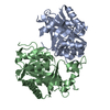
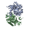
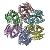
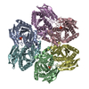
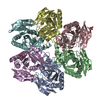
| Title | X-ray structure of the unliganded uridine phosphorylase from salmonella typhimurium in homodimeric form at 1.8A | ||||||
 Components Components | Uridine phosphorylase | ||||||
 Keywords Keywords | TRANSFERASE / Glycosyltransferase | ||||||
| Function / homology |  Function and homology information Function and homology informationnucleoside catabolic process / uridine phosphorylase / nucleotide catabolic process / UMP salvage / uridine phosphorylase activity / cytosolSimilarity search - Function | ||||||
| Biological species |  Salmonella typhimurium (bacteria) Salmonella typhimurium (bacteria) | ||||||
| Method | X-RAY DIFFRACTION / SYNCHROTRON / MOLECULAR REPLACEMENT / Resolution: 1.8 Å | ||||||
 Authors Authors | Mikhailov, A.M. / Lashkov, A.A. | ||||||
 Citation Citation | Journal: To be Published Title: X-ray structure of the unliganded uridine phosphorylase from salmonella typhimurium in homodimeric form at 1.8A Authors: Lashkov, A.A. / Mikhailov, A.M. | ||||||
| History |
|
- Structure visualization
| Structure viewer | Molecule: MolmilJmol/JSmol |
|---|
- Downloads & links
-Download
| PDBx/mmCIF format | 3dps.cif.gz | 107.1 KB | Display | PDBx/mmCIF format |
|---|---|---|---|---|
| PDB format | pdb3dps.ent.gz | 81.6 KB | Display | PDB format |
| PDBx/mmJSON format | 3dps.json.gz | Tree view | PDBx/mmJSON format | |
| Others |  Other downloads Other downloads |
-Validation report
| Arichive directory | https://data.pdbj.org/pub/pdb/validation_reports/dp/3dpsftp://data.pdbj.org/pub/pdb/validation_reports/dp/3dps | HTTPS FTP |
|---|
-Related structure data
| Related structure data |  2oxfS S: Starting model for refinement |
|---|---|
| Similar structure data |


-Links
 PDBj
PDBj- Assembly
| Deposited unit | 
| ||||||||
|---|---|---|---|---|---|---|---|---|---|
| 1 | 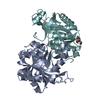
| ||||||||
| Unit cell |
| ||||||||
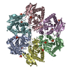
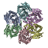
| Details | The biological assembly is a hexamer generated from the dimer in the asymmetric unit by the operations: 1-y, x-y, z and 1-x+y, x-1, z. |
-Components
| #1: Protein | / UrdPase / UPase Mass: 27169.092 Da / Num. of mol.: 2 Source method: isolated from a genetically manipulated source Source: (gene. exp.) Salmonella typhimurium (bacteria) / Strain: LT2 / Gene: udp / Plasmid: PBLUESCRIPT IISK / Production host: Escherichia coli (E. coli) / Strain (production host): Bl21 De3 / References: UniProt: P0A1F6, uridine phosphorylase#2: Chemical | ChemComp-GOL / | Glycerol  Mass: 92.094 Da / Num. of mol.: 1 / Source method: obtained synthetically / Formula: C3H8O3 Mass: 92.094 Da / Num. of mol.: 1 / Source method: obtained synthetically / Formula: C3H8O3#3: Water | ChemComp-HOH / | Water Mass: 18.015 Da / Num. of mol.: 254 / Source method: isolated from a natural source / Formula: H2O Mass: 18.015 Da / Num. of mol.: 254 / Source method: isolated from a natural source / Formula: H2O |
|---|
-Experimental details
-Experiment
| Experiment | Method: X-RAY DIFFRACTION / Number of used crystals: 1 |
|---|
- Sample preparation
| Crystal | Density Matthews: 1.93 Å3/Da / Density % sol: 36.21 % |
|---|---|
| Crystal grow | Temperature: 295 K / Method: vapor diffusion, hanging drop / pH: 5.2 Details: PEG 400, NaN3, GLYCEROL, pH 5.2, VAPOR DIFFUSION, HANGING DROP, temperature 295K |
-Data collection
| Diffraction | Mean temperature: 100 K |
|---|---|
| Diffraction source | Source: SYNCHROTRON / Site: EMBL/DESY, HAMBURG  / Beamline: X13 / Wavelength: 0.813 Å / Beamline: X13 / Wavelength: 0.813 Å |
| Detector | Type: MAR CCD 165 mm / Detector: CCD / Date: Nov 23, 2007 |
| Radiation | Monochromator: GAPHITE / Protocol: SINGLE WAVELENGTH / Monochromatic (M) / Laue (L): M / Scattering type: x-ray |
| Radiation wavelength | Wavelength: 0.813 Å / Relative weight: 1 |
| Reflection | Resolution: 1.8→20 Å / Num. all: 37630 / Num. obs: 37497 / % possible obs: 99.7 % / Observed criterion σ(F): 0 / Observed criterion σ(I): -3 / Biso Wilson estimate: 19.14 Å2 / Rmerge(I) obs: 0.058 |
- Processing
| Software |
| ||||||||||||||||||||||||||||||||||||||||||||||||||||||||||||||||||||||||||||||||||||||||||||||||||
|---|---|---|---|---|---|---|---|---|---|---|---|---|---|---|---|---|---|---|---|---|---|---|---|---|---|---|---|---|---|---|---|---|---|---|---|---|---|---|---|---|---|---|---|---|---|---|---|---|---|---|---|---|---|---|---|---|---|---|---|---|---|---|---|---|---|---|---|---|---|---|---|---|---|---|---|---|---|---|---|---|---|---|---|---|---|---|---|---|---|---|---|---|---|---|---|---|---|---|---|
| Refinement | Method to determine structure: MOLECULAR REPLACEMENT Starting model: PDB ENTRY 2OXF Resolution: 1.8→18.696 Å / Occupancy max: 1 / Occupancy min: 0.5 / SU ML: 0.21 / σ(F): 2.01 / Phase error: 18.18 / Stereochemistry target values: ML
| ||||||||||||||||||||||||||||||||||||||||||||||||||||||||||||||||||||||||||||||||||||||||||||||||||
| Solvent computation | Shrinkage radii: 0.9 Å / VDW probe radii: 1.11 Å / Solvent model: FLAT BULK SOLVENT MODEL / Bsol: 51.232 Å2 / ksol: 0.378 e/Å3 | ||||||||||||||||||||||||||||||||||||||||||||||||||||||||||||||||||||||||||||||||||||||||||||||||||
| Displacement parameters | Biso max: 162.77 Å2 / Biso mean: 25.463 Å2 / Biso min: 7.69 Å2
| ||||||||||||||||||||||||||||||||||||||||||||||||||||||||||||||||||||||||||||||||||||||||||||||||||
| Refinement step | Cycle: LAST / Resolution: 1.8→18.696 Å
| ||||||||||||||||||||||||||||||||||||||||||||||||||||||||||||||||||||||||||||||||||||||||||||||||||
| Refine LS restraints |
| ||||||||||||||||||||||||||||||||||||||||||||||||||||||||||||||||||||||||||||||||||||||||||||||||||
| LS refinement shell | Refine-ID: X-RAY DIFFRACTION / Total num. of bins used: 13
|
