 Movie
Movie Controller

[English] 日本語
 Yorodumi
- PDB-2iq5: Unliganded Crystal Structure of the Uridine Phosphorylase from Sa... -
+ Open data

- Basic information
| Entry | Database: PDB / ID: 2iq5 | ||||||
|---|---|---|---|---|---|---|---|
| Title | Unliganded Crystal Structure of the Uridine Phosphorylase from Salmonella Typhimurium at 1.90 A Resolution | ||||||
 Components Components | Uridine phosphorylase | ||||||
 Keywords Keywords | TRANSFERASE / NUCLEOSIDE PHOSPHORYLASE | ||||||
| Function / homology |  Function and homology information Function and homology informationnucleoside catabolic process / uridine phosphorylase / nucleotide catabolic process / UMP salvage / uridine phosphorylase activity / cytosolSimilarity search - Function | ||||||
| Biological species |  Salmonella typhimurium (bacteria) Salmonella typhimurium (bacteria) | ||||||
| Method | X-RAY DIFFRACTION / SYNCHROTRON / MOLECULAR REPLACEMENT / Resolution: 1.9 Å | ||||||
 Authors Authors | Timofeev, V.I. / Dontsova, M.V. / Gabdoulkhakov, A.G. / Pavlyuk, B.P. / Mikhailov, A.M. | ||||||
 Citation Citation | Journal: To be Published Title: Unliganded Crystal Structure of the Uridine Phosphorylase from Salmonella Typhimurium at 1.90 A Resolution Authors: Timofeev, V.I. / Dontsova, M.V. / Gabdoulkhakov, A.G. / Lashkov, A.A. / Voelter, V. / Kachalova, G.S. / Pavlyuk, B.P. / Mikhailov, A.M. | ||||||
| History |
|
- Structure visualization
| Structure viewer | Molecule: MolmilJmol/JSmol |
|---|
- Downloads & links
-Download
| PDBx/mmCIF format | 2iq5.cif.gz | 181.7 KB | Display | PDBx/mmCIF format |
|---|---|---|---|---|
| PDB format | pdb2iq5.ent.gz | 145.2 KB | Display | PDB format |
| PDBx/mmJSON format | 2iq5.json.gz | Tree view | PDBx/mmJSON format | |
| Others |  Other downloads Other downloads |
-Validation report
| Arichive directory | https://data.pdbj.org/pub/pdb/validation_reports/iq/2iq5ftp://data.pdbj.org/pub/pdb/validation_reports/iq/2iq5 | HTTPS FTP |
|---|
-Related structure data
| Similar structure data |
|---|


-Links
 PDBj
PDBj- Assembly
| Deposited unit | 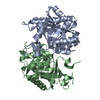
| ||||||||
|---|---|---|---|---|---|---|---|---|---|
| 1 | 
| ||||||||
| Unit cell |
| ||||||||
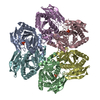
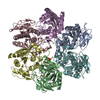
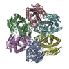
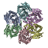
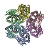
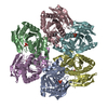
| Details | The biological assembly is a hexamer generated from the dimer, which presented in the asymmetric unit. |
-Components
| #1: Protein | / UrdPase / UPase Mass: 27037.896 Da / Num. of mol.: 2 Source method: isolated from a genetically manipulated source Source: (gene. exp.) Salmonella typhimurium (bacteria) / Strain: LT2 / Gene: UDP / Plasmid: PBLUESCRIPT IISK / Species (production host): Escherichia coli / Production host: Escherichia coli BL21(DE3) (bacteria) / Strain (production host): BL21 DE3 / References: UniProt: P0A1F6, uridine phosphorylase#2: Water | ChemComp-HOH / | Water Mass: 18.015 Da / Num. of mol.: 175 / Source method: isolated from a natural source / Formula: H2O Mass: 18.015 Da / Num. of mol.: 175 / Source method: isolated from a natural source / Formula: H2O |
|---|
-Experimental details
-Experiment
| Experiment | Method: X-RAY DIFFRACTION / Number of used crystals: 1 |
|---|
- Sample preparation
| Crystal | Density Matthews: 1.96 Å3/Da / Density % sol: 37.27 % |
|---|---|
| Crystal grow | Temperature: 297 K / Method: vapor diffusion, hanging drop / Details: VAPOR DIFFUSION, HANGING DROP, temperature 297K |
-Data collection
| Diffraction | Mean temperature: 100 K |
|---|---|
| Diffraction source | Source: SYNCHROTRON / Site: EMBL/DESY, HAMBURG  / Beamline: X11 / Wavelength: 0.8126 / Beamline: X11 / Wavelength: 0.8126 |
| Detector | Type: MAR CCD 165 mm / Detector: CCD / Date: Nov 15, 2005 / Details: mirrors |
| Radiation | Monochromator: GRAPHITE / Protocol: SINGLE WAVELENGTH / Monochromatic (M) / Laue (L): M / Scattering type: x-ray |
| Radiation wavelength | Wavelength: 0.8126 Å / Relative weight: 1 |
| Reflection | Resolution: 1.9→76.7 Å / Num. obs: 32175 / Redundancy: 2.71 % |
| Reflection shell | Resolution: 1.9→1.95 Å |
- Processing
| Software | Name: REFMAC / Version: 5.2.0003 / Classification: refinement | ||||||||||||||||||||||||||||||||||||||||||||||||||||||||||||||||||||||||||||||||||||||||||||||||||||||||||||||||||||||||||||||||||||||||||||||||||||||||||||||||||||||||||
|---|---|---|---|---|---|---|---|---|---|---|---|---|---|---|---|---|---|---|---|---|---|---|---|---|---|---|---|---|---|---|---|---|---|---|---|---|---|---|---|---|---|---|---|---|---|---|---|---|---|---|---|---|---|---|---|---|---|---|---|---|---|---|---|---|---|---|---|---|---|---|---|---|---|---|---|---|---|---|---|---|---|---|---|---|---|---|---|---|---|---|---|---|---|---|---|---|---|---|---|---|---|---|---|---|---|---|---|---|---|---|---|---|---|---|---|---|---|---|---|---|---|---|---|---|---|---|---|---|---|---|---|---|---|---|---|---|---|---|---|---|---|---|---|---|---|---|---|---|---|---|---|---|---|---|---|---|---|---|---|---|---|---|---|---|---|---|---|---|---|---|---|
| Refinement | Method to determine structure: MOLECULAR REPLACEMENT / Resolution: 1.9→10 Å / Cor.coef. Fo:Fc: 0.953 / Cor.coef. Fo:Fc free: 0.927 / SU B: 10.2 / SU ML: 0.135 / Cross valid method: THROUGHOUT / ESU R: 0.173 / ESU R Free: 0.168 / Stereochemistry target values: MAXIMUM LIKELIHOOD / Details: HYDROGENS HAVE BEEN ADDED IN THE RIDING POSITIONS
| ||||||||||||||||||||||||||||||||||||||||||||||||||||||||||||||||||||||||||||||||||||||||||||||||||||||||||||||||||||||||||||||||||||||||||||||||||||||||||||||||||||||||||
| Solvent computation | Ion probe radii: 0.8 Å / Shrinkage radii: 0.8 Å / VDW probe radii: 1.4 Å / Solvent model: MASK | ||||||||||||||||||||||||||||||||||||||||||||||||||||||||||||||||||||||||||||||||||||||||||||||||||||||||||||||||||||||||||||||||||||||||||||||||||||||||||||||||||||||||||
| Displacement parameters | Biso mean: 32.893 Å2
| ||||||||||||||||||||||||||||||||||||||||||||||||||||||||||||||||||||||||||||||||||||||||||||||||||||||||||||||||||||||||||||||||||||||||||||||||||||||||||||||||||||||||||
| Refinement step | Cycle: LAST / Resolution: 1.9→10 Å
| ||||||||||||||||||||||||||||||||||||||||||||||||||||||||||||||||||||||||||||||||||||||||||||||||||||||||||||||||||||||||||||||||||||||||||||||||||||||||||||||||||||||||||
| Refine LS restraints |
| ||||||||||||||||||||||||||||||||||||||||||||||||||||||||||||||||||||||||||||||||||||||||||||||||||||||||||||||||||||||||||||||||||||||||||||||||||||||||||||||||||||||||||
| LS refinement shell | Resolution: 1.9→1.95 Å / Total num. of bins used: 20
|
