 Movie
Movie Controller

[English] 日本語
 Yorodumi
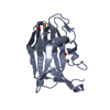
- PDB-4lbf: Crystal structure of HUMAN ALPHA-DEFENSIN 1 (HNP1) I20A/L25A mutant -
+ Open data

- Basic information
| Entry | Database: PDB / ID: 4lbf | ||||||
|---|---|---|---|---|---|---|---|
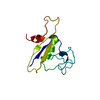
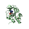
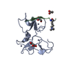
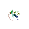
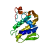
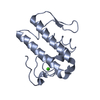
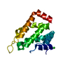
| Title | Crystal structure of HUMAN ALPHA-DEFENSIN 1 (HNP1) I20A/L25A mutant | ||||||
 Components Components | Neutrophil defensin 1 | ||||||
 Keywords Keywords |  ANTIMICROBIAL PROTEIN / ANTIMICROBIAL PEPTIDE / HUMAN ALPHA DEFENSIN 1 / HUMAN NEUTROPHIL PEPTIDE 1 / ANTIBIOTIC / ANTIVIRAL DEFENSE / DEFENSIN / DISULFIDE BOND / FUNGICIDE / PHOSPHOPROTEIN / SECRETED ANTIMICROBIAL PROTEIN / ANTIMICROBIAL PEPTIDE / HUMAN ALPHA DEFENSIN 1 / HUMAN NEUTROPHIL PEPTIDE 1 / ANTIBIOTIC / ANTIVIRAL DEFENSE / DEFENSIN / DISULFIDE BOND / FUNGICIDE / PHOSPHOPROTEIN / SECRETED | ||||||
| Function / homology |  Function and homology information Function and homology informationpore-forming activity / Defensins / disruption of plasma membrane integrity in another organism / killing by host of symbiont cells / T cell chemotaxis / Alpha-defensins / defense response to protozoan / intracellular estrogen receptor signaling pathway / defense response to fungus / innate immune response in mucosa ...pore-forming activity / Defensins / disruption of plasma membrane integrity in another organism / killing by host of symbiont cells / T cell chemotaxis / Alpha-defensins / defense response to protozoan / intracellular estrogen receptor signaling pathway / defense response to fungus / innate immune response in mucosa / Golgi lumen / chemotaxis / azurophil granule lumen / antimicrobial humoral immune response mediated by antimicrobial peptide / antibacterial humoral response / collagen-containing extracellular matrix / defense response to virus / killing of cells of another organism / cellular response to lipopolysaccharide / defense response to Gram-negative bacterium / defense response to Gram-positive bacterium / immune response / Neutrophil degranulation / extracellular space / extracellular exosome / extracellular regionSimilarity search - Function | ||||||
| Biological species |  Homo sapiens (human) Homo sapiens (human) | ||||||
| Method | X-RAY DIFFRACTION / MOLECULAR REPLACEMENT / Resolution: 1.7 Å | ||||||
 Authors Authors | Tolbert, W.D. / Wu, X. / Pazgier, M. | ||||||
 Citation Citation | Journal: Plos One / Year: 2013 Title: Single, Double and Quadruple Alanine Substitutions at Oligomeric Interfaces Identify Hydrophobicity as the Key Determinant of Human Neutrophil Alpha Defensin HNP1 Function. Authors: Zhao, L. / Tolbert, W.D. / Ericksen, B. / Zhan, C. / Wu, X. / Yuan, W. / Li, X. / Pazgier, M. / Lu, W. | ||||||
| History |
|
- Structure visualization
| Structure viewer | Molecule: MolmilJmol/JSmol |
|---|
- Downloads & links
-Download
| PDBx/mmCIF format | 4lbf.cif.gz | 59.6 KB | Display | PDBx/mmCIF format |
|---|---|---|---|---|
| PDB format | pdb4lbf.ent.gz | 46.1 KB | Display | PDB format |
| PDBx/mmJSON format | 4lbf.json.gz | Tree view | PDBx/mmJSON format | |
| Others |  Other downloads Other downloads |
-Validation report
| Arichive directory | https://data.pdbj.org/pub/pdb/validation_reports/lb/4lbfftp://data.pdbj.org/pub/pdb/validation_reports/lb/4lbf | HTTPS FTP |
|---|
-Related structure data
| Related structure data | 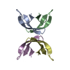 4lb1C 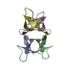 4lb7C 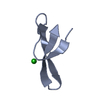 4lbbC  3gnyS S: Starting model for refinement C: citing same article ( |
|---|---|
| Similar structure data |
-Links
 PDBj
PDBj


- Assembly
| Deposited unit | 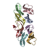
| ||||||||
|---|---|---|---|---|---|---|---|---|---|
| 1 | 
| ||||||||
| 2 | 
| ||||||||
| Unit cell |
|
-Components
| #1: Protein/peptide | Mass: 3367.951 Da / Num. of mol.: 8 / Mutation: I20A, L25A / Source method: obtained synthetically / Source: (synth.) Homo sapiens (human) / References: UniProt: P59665#2: Chemical | ChemComp-GOL / | Glycerol  Mass: 92.094 Da / Num. of mol.: 1 / Source method: obtained synthetically / Formula: C3H8O3 Mass: 92.094 Da / Num. of mol.: 1 / Source method: obtained synthetically / Formula: C3H8O3#3: Water | ChemComp-HOH / | Water Mass: 18.015 Da / Num. of mol.: 209 / Source method: isolated from a natural source / Formula: H2O Mass: 18.015 Da / Num. of mol.: 209 / Source method: isolated from a natural source / Formula: H2O |
|---|
-Experimental details
-Experiment
| Experiment | Method: X-RAY DIFFRACTION / Number of used crystals: 1 |
|---|
- Sample preparation
| Crystal | Density Matthews: 1.82 Å3/Da / Density % sol: 32.58 % |
|---|---|
| Crystal grow | Temperature: 298 K / Method: vapor diffusion, hanging drop / pH: 8.5 Details: 2 M ammonium sulfate, 0.1 M Tris-HCl pH 8.5, VAPOR DIFFUSION, HANGING DROP, temperature 298.0K |
-Data collection
| Diffraction | Mean temperature: 100 K |
|---|---|
| Diffraction source | Source: ROTATING ANODE / Type: RIGAKU MICROMAX-007 / Wavelength: 1.54 Å |
| Detector | Type: RIGAKU RAXIS IV / Detector: IMAGE PLATE / Date: May 27, 2009 / Details: CONFOCAL |
| Radiation | Monochromator: NONE / Protocol: SINGLE WAVELENGTH / Monochromatic (M) / Laue (L): M / Scattering type: x-ray |
| Radiation wavelength | Wavelength: 1.54 Å / Relative weight: 1 |
| Reflection | Resolution: 1.66→50 Å / Num. all: 22854 / Num. obs: 22465 / % possible obs: 98.3 % / Observed criterion σ(F): 0 / Observed criterion σ(I): 0 / Redundancy: 2.3 % / Rmerge(I) obs: 0.081 / Net I/σ(I): 16.5 |
| Reflection shell | Resolution: 1.66→1.69 Å / Redundancy: 3.3 % / Rmerge(I) obs: 0.448 / Mean I/σ(I) obs: 1.6 / % possible all: 84.1 |
- Processing
| Software |
| ||||||||||||||||||||||||||||||||||||||||||||||||||||||||||||||||||||||||||||||||||||||||||||||||||||||||||||||||||||||||||||||||||||||||||||||||||||||||||||||||||||||||||||||||||||||
|---|---|---|---|---|---|---|---|---|---|---|---|---|---|---|---|---|---|---|---|---|---|---|---|---|---|---|---|---|---|---|---|---|---|---|---|---|---|---|---|---|---|---|---|---|---|---|---|---|---|---|---|---|---|---|---|---|---|---|---|---|---|---|---|---|---|---|---|---|---|---|---|---|---|---|---|---|---|---|---|---|---|---|---|---|---|---|---|---|---|---|---|---|---|---|---|---|---|---|---|---|---|---|---|---|---|---|---|---|---|---|---|---|---|---|---|---|---|---|---|---|---|---|---|---|---|---|---|---|---|---|---|---|---|---|---|---|---|---|---|---|---|---|---|---|---|---|---|---|---|---|---|---|---|---|---|---|---|---|---|---|---|---|---|---|---|---|---|---|---|---|---|---|---|---|---|---|---|---|---|---|---|---|---|
| Refinement | Method to determine structure: MOLECULAR REPLACEMENT Starting model: PDB entry 3GNY Resolution: 1.7→20 Å / Cor.coef. Fo:Fc: 0.964 / Cor.coef. Fo:Fc free: 0.955 / SU B: 2.857 / SU ML: 0.093 / Cross valid method: THROUGHOUT / σ(F): 0 / ESU R: 0.132 / ESU R Free: 0.127 / Stereochemistry target values: MAXIMUM LIKELIHOOD
| ||||||||||||||||||||||||||||||||||||||||||||||||||||||||||||||||||||||||||||||||||||||||||||||||||||||||||||||||||||||||||||||||||||||||||||||||||||||||||||||||||||||||||||||||||||||
| Solvent computation | Ion probe radii: 0.8 Å / Shrinkage radii: 0.8 Å / VDW probe radii: 1.4 Å / Solvent model: BABINET MODEL WITH MASK | ||||||||||||||||||||||||||||||||||||||||||||||||||||||||||||||||||||||||||||||||||||||||||||||||||||||||||||||||||||||||||||||||||||||||||||||||||||||||||||||||||||||||||||||||||||||
| Displacement parameters | Biso mean: 29.513 Å2
| ||||||||||||||||||||||||||||||||||||||||||||||||||||||||||||||||||||||||||||||||||||||||||||||||||||||||||||||||||||||||||||||||||||||||||||||||||||||||||||||||||||||||||||||||||||||
| Refinement step | Cycle: LAST / Resolution: 1.7→20 Å
| ||||||||||||||||||||||||||||||||||||||||||||||||||||||||||||||||||||||||||||||||||||||||||||||||||||||||||||||||||||||||||||||||||||||||||||||||||||||||||||||||||||||||||||||||||||||
| Refine LS restraints |
|