[English] 日本語
Yorodumi
- PDB-4kng: Crystal structure of human LGR5-RSPO1-RNF43 -

+
Open data


ID or keywords:

Loading...

-
Basic information

Entry
Database: PDB / ID: 4kng
TitleCrystal structure of human LGR5-RSPO1-RNF43
Components
  • E3 ubiquitin-protein ligase RNF43
  • Leucine-rich repeat-containing G-protein coupled receptor 5
  • R-spondin-1
KeywordsSignaling Protein / Membrane Protein / Leucine-rich repeat / cysteine-rich domain / furin-repeat / protease-associated domain / ligand recognition / protein-protein interaction / N-linked glycosylation
Function / homology
Function and homology information


oocyte differentiation / epithelial cell proliferation involved in renal tubule morphogenesis / Wnt receptor catabolic process / protein-hormone receptor activity / Signaling by RNF43 mutants / regulation of receptor internalization / multicellular organism development / G protein-coupled peptide receptor activity / frizzled binding / negative regulation of Wnt signaling pathway ...oocyte differentiation / epithelial cell proliferation involved in renal tubule morphogenesis / Wnt receptor catabolic process / protein-hormone receptor activity / Signaling by RNF43 mutants / regulation of receptor internalization / multicellular organism development / G protein-coupled peptide receptor activity / frizzled binding / negative regulation of Wnt signaling pathway / plasma membrane => GO:0005886 / inner ear development / positive regulation of Wnt signaling pathway / hair follicle development / Regulation of FZD by ubiquitination / trans-Golgi network membrane / stem cell proliferation / G protein-coupled receptor binding / G protein-coupled receptor activity / RING-type E3 ubiquitin transferase / adenylate cyclase-activating G protein-coupled receptor signaling pathway / Wnt signaling pathway / ubiquitin-protein transferase activity / positive regulation of canonical Wnt signaling pathway / ubiquitin protein ligase activity / transmembrane signaling receptor activity / heparin binding / nuclear envelope / regulation of cell population proliferation / ubiquitin-dependent protein catabolic process / protein ubiquitination / positive regulation of protein phosphorylation / G protein-coupled receptor signaling pathway / signaling receptor binding / endoplasmic reticulum membrane / Golgi apparatus / extracellular region / nucleoplasm / metal ion binding / nucleus / plasma membrane
Similarity search - Function
ZNRF-3, ectodomain / ZNRF-3 Ectodomain / R-spondin, Fu-CRD domain / Furin-like repeat, cysteine-rich / Glycoprotein hormone receptor family / Hormone Receptor, Insulin-like Growth Factor Receptor 1; Chain A domain 2 / Hormone Receptor, Insulin-like Growth Factor Receptor 1; Chain A, domain 2 / Leucine rich repeat N-terminal domain / Ring finger domain / Glucose Oxidase; domain 1 - #30 ...ZNRF-3, ectodomain / ZNRF-3 Ectodomain / R-spondin, Fu-CRD domain / Furin-like repeat, cysteine-rich / Glycoprotein hormone receptor family / Hormone Receptor, Insulin-like Growth Factor Receptor 1; Chain A domain 2 / Hormone Receptor, Insulin-like Growth Factor Receptor 1; Chain A, domain 2 / Leucine rich repeat N-terminal domain / Ring finger domain / Glucose Oxidase; domain 1 - #30 / Leucine-rich repeat N-terminal domain / Leucine rich repeat N-terminal domain / Glucose Oxidase; domain 1 / Leucine-rich repeats, bacterial type / Thrombospondin type-1 (TSP1) repeat superfamily / Thrombospondin type-1 (TSP1) repeat profile. / Thrombospondin type 1 repeats / Thrombospondin type-1 (TSP1) repeat / Leucine Rich Repeat / Leucine-rich repeat, SDS22-like subfamily / Leucine-rich repeat, LRR (right-handed beta-alpha superhelix) / Ribonuclease Inhibitor / Alpha-Beta Horseshoe / Furin-like repeat / Furin-like repeats / Leucine rich repeat / Leucine-rich repeat, typical subtype / Leucine-rich repeats, typical (most populated) subfamily / Leucine-rich repeat profile. / Ring finger / Growth factor receptor cysteine-rich domain superfamily / Leucine-rich repeat / Leucine-rich repeat domain superfamily / Zinc finger RING-type profile. / Zinc finger, RING-type / 3-Layer(bba) Sandwich / Ribbon / G protein-coupled receptor, rhodopsin-like / GPCR, rhodopsin-like, 7TM / G-protein coupled receptors family 1 profile. / 7 transmembrane receptor (rhodopsin family) / Zinc finger, RING/FYVE/PHD-type / Mainly Beta / Alpha Beta
Similarity search - Domain/homology
NICKEL (II) ION / Leucine-rich repeat-containing G-protein coupled receptor 5 / R-spondin-1 / E3 ubiquitin-protein ligase RNF43
Similarity search - Component
Biological speciesHomo sapiens (human)
MethodX-RAY DIFFRACTION / SYNCHROTRON / SIRAS / Resolution: 2.5 Å
AuthorsChen, P.H. / He, X.
CitationJournal: Genes Dev. / Year: 2013
Title: The structural basis of R-spondin recognition by LGR5 and RNF43.
Authors: Chen, P.H. / Chen, X. / Lin, Z. / Fang, D. / He, X.
History
DepositionMay 9, 2013Deposition site: RCSB / Processing site: RCSB
Revision 1.0Jun 19, 2013Provider: repository / Type: Initial release
Revision 1.1Jul 3, 2013Group: Data collection
Revision 1.2Jul 24, 2013Group: Database references
Revision 1.3Jul 29, 2020Group: Data collection / Database references ...Data collection / Database references / Derived calculations / Structure summary
Category: chem_comp / entity ...chem_comp / entity / pdbx_chem_comp_identifier / pdbx_entity_nonpoly / pdbx_struct_conn_angle / struct_conn / struct_ref_seq_dif / struct_site / struct_site_gen
Item: _chem_comp.name / _chem_comp.type ..._chem_comp.name / _chem_comp.type / _entity.pdbx_description / _pdbx_entity_nonpoly.name / _pdbx_struct_conn_angle.ptnr1_auth_asym_id / _pdbx_struct_conn_angle.ptnr1_label_asym_id / _pdbx_struct_conn_angle.ptnr3_auth_asym_id / _pdbx_struct_conn_angle.ptnr3_label_asym_id / _pdbx_struct_conn_angle.value / _struct_conn.pdbx_dist_value / _struct_conn.pdbx_leaving_atom_flag / _struct_conn.pdbx_role / _struct_conn.ptnr1_auth_asym_id / _struct_conn.ptnr1_auth_seq_id / _struct_conn.ptnr1_label_asym_id / _struct_conn.ptnr1_label_seq_id / _struct_conn.ptnr2_auth_asym_id / _struct_conn.ptnr2_auth_seq_id / _struct_conn.ptnr2_label_asym_id / _struct_conn.ptnr2_label_seq_id / _struct_ref_seq_dif.details
Description: Carbohydrate remediation / Provider: repository / Type: Remediation

-
Structure visualization

Structure viewerMolecule:
MolmilJmol/JSmol

Downloads & links

-
Assembly

Deposited unit
A: Leucine-rich repeat-containing G-protein coupled receptor 5
B: Leucine-rich repeat-containing G-protein coupled receptor 5
M: R-spondin-1
P: R-spondin-1
E: E3 ubiquitin-protein ligase RNF43
F: E3 ubiquitin-protein ligase RNF43
hetero molecules


Theoretical massNumber of molelcules
Total (without water)178,8839
Polymers178,3826
Non-polymers5013
Water3,693205
1


  • Idetical with deposited unit
  • defined by author
TypeNameSymmetry operationNumber
identity operation1_555x,y,z1
Unit cell
Length a, b, c (Å)104.575, 120.971, 181.009
Angle α, β, γ (deg.)90.00, 90.00, 90.00
Int Tables number19
Space group name H-MP212121
Noncrystallographic symmetry (NCS)NCS domain:
IDEns-IDDetails
11A
21B
12M
22P
13E
23F

NCS domain segments:
Dom-IDComponent-IDEns-IDRefine codeAuth asym-IDAuth seq-ID
1010A30 - 542
2010B30 - 542
1020M40 - 132
2020P40 - 132
1030E44 - 190
2030F44 - 190

NCS ensembles :
ID
1
2
3
Details0.009563 0.000000 0.000000 0.00000 0.000000 0.008266 0.000000 0.00000 0.000000 0.000000 0.005525 0.00000

-
Components

-
Protein , 3 types, 6 molecules ABMPEF

#1: Protein Leucine-rich repeat-containing G-protein coupled receptor 5 / G-protein coupled receptor 49 / G-protein coupled receptor 67 / G-protein coupled receptor HG38


Mass: 59278.367 Da / Num. of mol.: 2 / Fragment: extracellular domain
Source method: isolated from a genetically manipulated source
Source: (gene. exp.) Homo sapiens (human) / Gene: LGR5, GPR49, GPR67 / Plasmid: PVLAD6 / Cell line (production host): HEK293 / Production host: Homo sapiens (human) / References: UniProt: O75473
#2: Protein R-spondin-1 / Roof plate-specific spondin-1 / hRspo1


Mass: 12362.315 Da / Num. of mol.: 2 / Fragment: furin repeats
Source method: isolated from a genetically manipulated source
Source: (gene. exp.) Homo sapiens (human) / Gene: RSPO1 / Plasmid: PVLAD6 / Cell line (production host): HEK293 / Production host: Homo sapiens (human) / References: UniProt: Q2MKA7
#3: Protein E3 ubiquitin-protein ligase RNF43 / RING finger protein 43


Mass: 17550.203 Da / Num. of mol.: 2 / Fragment: PA domain
Source method: isolated from a genetically manipulated source
Source: (gene. exp.) Homo sapiens (human) / Gene: RNF43 / Plasmid: PVLAD6 / Cell line (production host): HEK293 / Production host: Homo sapiens (human)
References: UniProt: Q68DV7, Ligases; Forming carbon-nitrogen bonds; Acid-amino-acid ligases (peptide synthases)

-
Sugars , 1 types, 2 molecules

#4: Sugar ChemComp-NAG / 2-acetamido-2-deoxy-beta-D-glucopyranose / N-Acetylglucosamine


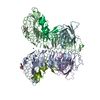
Type: D-saccharide, beta linking / Mass: 221.208 Da / Num. of mol.: 2
Source method: isolated from a genetically manipulated source
Formula: C8H15NO6
IdentifierTypeProgram
DGlcpNAcbCONDENSED IUPAC CARBOHYDRATE SYMBOLGMML 1.0
N-acetyl-b-D-glucopyranosamineCOMMON NAMEGMML 1.0
b-D-GlcpNAcIUPAC CARBOHYDRATE SYMBOLPDB-CARE 1.0
GlcNAcSNFG CARBOHYDRATE SYMBOLGMML 1.0

-
Non-polymers , 2 types, 206 molecules

#5: Chemical ChemComp-NI / NICKEL (II) ION / Nickel


Mass: 58.693 Da / Num. of mol.: 1 / Source method: obtained synthetically / Formula: Ni
#6: Water ChemComp-HOH / water / Water


Mass: 18.015 Da / Num. of mol.: 205 / Source method: isolated from a natural source / Formula: H2O

-
Experimental details

-
Experiment

ExperimentMethod: X-RAY DIFFRACTION / Number of used crystals: 1

-
Sample preparation

CrystalDensity Matthews: 3.21 Å3/Da / Density % sol: 61.67 %
Crystal growTemperature: 293 K / Method: vapor diffusion, sitting drop / pH: 8
Details: 10% PEG4000, 0.2M ammonium sulfate, 7% Sucrose and 0.1 M TRIS pH 8.0, VAPOR DIFFUSION, SITTING DROP, temperature 293K

-
Data collection

DiffractionMean temperature: 100 K
Diffraction sourceSource: SYNCHROTRON / Site: APS / Beamline: 21-ID-D / Wavelength: 0.979 Å
DetectorType: MARRESEARCH / Detector: CCD / Date: Oct 15, 2012
RadiationMonochromator: Ni / Protocol: MAD / Monochromatic (M) / Laue (L): M / Scattering type: x-ray
Radiation wavelengthWavelength: 0.979 Å / Relative weight: 1
ReflectionResolution: 2.5→39.51 Å / Num. all: 74020 / % possible obs: 97.1 % / Observed criterion σ(F): 0 / Observed criterion σ(I): 0

-
Processing

Software
NameVersionClassification
HKL-3000data collection
X-PLORmodel building
REFMAC5.7.0029refinement
HKL-3000data reduction
SCALAdata scaling
X-PLORphasing
RefinementMethod to determine structure: SIRAS / Resolution: 2.5→39.51 Å / Cor.coef. Fo:Fc: 0.93 / Cor.coef. Fo:Fc free: 0.904 / SU B: 11.435 / SU ML: 0.246 / Cross valid method: THROUGHOUT / ESU R: 0.371 / ESU R Free: 0.276 / Stereochemistry target values: MAXIMUM LIKELIHOOD / Details: HYDROGENS HAVE BEEN ADDED IN THE RIDING POSITIONS
RfactorNum. reflection% reflectionSelection details
Rfree0.27633 3894 5 %RANDOM
Rwork0.23197 ---
obs0.23425 74020 97.08 %-
all-74020 --
Solvent computationIon probe radii: 0.8 Å / Shrinkage radii: 0.8 Å / VDW probe radii: 1.2 Å / Solvent model: MASK
Displacement parametersBiso mean: 76.177 Å2
Baniso -1Baniso -2Baniso -3
1--0.07 Å2-0 Å20 Å2
2--0.03 Å2-0 Å2
3---0.04 Å2
Refinement stepCycle: LAST / Resolution: 2.5→39.51 Å
ProteinNucleic acidLigandSolventTotal
Num. atoms11012 0 29 205 11246
Refine LS restraints
Refine-IDTypeDev idealDev ideal targetNumber
X-RAY DIFFRACTIONr_bond_refined_d0.0130.01911321
X-RAY DIFFRACTIONr_bond_other_d0.0040.0210961
X-RAY DIFFRACTIONr_angle_refined_deg1.8441.98415435
X-RAY DIFFRACTIONr_angle_other_deg1.1513.00425227
X-RAY DIFFRACTIONr_dihedral_angle_1_deg9.88251405
X-RAY DIFFRACTIONr_dihedral_angle_2_deg41.78424.534483
X-RAY DIFFRACTIONr_dihedral_angle_3_deg21.212151947
X-RAY DIFFRACTIONr_dihedral_angle_4_deg18.8111558
X-RAY DIFFRACTIONr_chiral_restr0.1020.21763
X-RAY DIFFRACTIONr_gen_planes_refined0.0080.02112681
X-RAY DIFFRACTIONr_gen_planes_other0.0040.022497
X-RAY DIFFRACTIONr_nbd_refined
X-RAY DIFFRACTIONr_nbd_other
X-RAY DIFFRACTIONr_nbtor_refined
X-RAY DIFFRACTIONr_nbtor_other
X-RAY DIFFRACTIONr_xyhbond_nbd_refined
X-RAY DIFFRACTIONr_xyhbond_nbd_other
X-RAY DIFFRACTIONr_metal_ion_refined
X-RAY DIFFRACTIONr_metal_ion_other
X-RAY DIFFRACTIONr_symmetry_vdw_refined
X-RAY DIFFRACTIONr_symmetry_vdw_other
X-RAY DIFFRACTIONr_symmetry_hbond_refined
X-RAY DIFFRACTIONr_symmetry_hbond_other
X-RAY DIFFRACTIONr_symmetry_metal_ion_refined
X-RAY DIFFRACTIONr_symmetry_metal_ion_other
X-RAY DIFFRACTIONr_mcbond_it5.6657.445647
X-RAY DIFFRACTIONr_mcbond_other5.6617.4395646
X-RAY DIFFRACTIONr_mcangle_it8.75611.1317043
X-RAY DIFFRACTIONr_scbond_it5.3627.8385674
X-RAY DIFFRACTIONr_scangle_it
X-RAY DIFFRACTIONr_rigid_bond_restr
X-RAY DIFFRACTIONr_sphericity_free
X-RAY DIFFRACTIONr_sphericity_bonded
Refine LS restraints NCS

Refine-ID: X-RAY DIFFRACTION / Type: interatomic distance / Weight position: 0.05

Ens-IDDom-IDAuth asym-IDNumberRms dev position (Å)
11A274560.14
12B274560.14
21M48110.12
22P48110.12
31E76890.18
32F76890.18
LS refinement shellResolution: 2.498→2.562 Å / Total num. of bins used: 20
RfactorNum. reflection% reflection
Rfree0.422 241 -
Rwork0.382 5121 -
obs--91.89 %

+
About Yorodumi

-
News

-
Feb 9, 2022. New format data for meta-information of EMDB entries

New format data for meta-information of EMDB entries

  • Version 3 of the EMDB header file is now the official format.
  • The previous official version 1.9 will be removed from the archive.

Related info.:EMDB header

External links:wwPDB to switch to version 3 of the EMDB data model

-
Aug 12, 2020. Covid-19 info

Covid-19 info

URL: https://pdbj.org/emnavi/covid19.php

New page: Covid-19 featured information page in EM Navigator.

Related info.:Covid-19 info / Mar 5, 2020. Novel coronavirus structure data

+
Mar 5, 2020. Novel coronavirus structure data

Novel coronavirus structure data

Related info.:Yorodumi Speices / Aug 12, 2020. Covid-19 info

External links:COVID-19 featured content - PDBj / Molecule of the Month (242):Coronavirus Proteases

+
Jan 31, 2019. EMDB accession codes are about to change! (news from PDBe EMDB page)

EMDB accession codes are about to change! (news from PDBe EMDB page)

  • The allocation of 4 digits for EMDB accession codes will soon come to an end. Whilst these codes will remain in use, new EMDB accession codes will include an additional digit and will expand incrementally as the available range of codes is exhausted. The current 4-digit format prefixed with “EMD-” (i.e. EMD-XXXX) will advance to a 5-digit format (i.e. EMD-XXXXX), and so on. It is currently estimated that the 4-digit codes will be depleted around Spring 2019, at which point the 5-digit format will come into force.
  • The EM Navigator/Yorodumi systems omit the EMD- prefix.

Related info.:Q: What is EMD? / ID/Accession-code notation in Yorodumi/EM Navigator

External links:EMDB Accession Codes are Changing Soon! / Contact to PDBj

+
Jul 12, 2017. Major update of PDB

Major update of PDB

  • wwPDB released updated PDB data conforming to the new PDBx/mmCIF dictionary.
  • This is a major update changing the version number from 4 to 5, and with Remediation, in which all the entries are updated.
  • In this update, many items about electron microscopy experimental information are reorganized (e.g. em_software).
  • Now, EM Navigator and Yorodumi are based on the updated data.

External links:wwPDB Remediation / Enriched Model Files Conforming to OneDep Data Standards Now Available in the PDB FTP Archive

-
Yorodumi

Thousand views of thousand structures

  • Yorodumi is a browser for structure data from EMDB, PDB, SASBDB, etc.
  • This page is also the successor to EM Navigator detail page, and also detail information page/front-end page for Omokage search.
  • The word "yorodu" (or yorozu) is an old Japanese word meaning "ten thousand". "mi" (miru) is to see.

Related info.:EMDB / PDB / SASBDB / Comparison of 3 databanks / Yorodumi Search / Aug 31, 2016. New EM Navigator & Yorodumi / Yorodumi Papers / Jmol/JSmol / Function and homology information / Changes in new EM Navigator and Yorodumi

Read more