 Movie
Movie Controller


+ Open data

- Basic information
| Entry | Database: PDB / ID: 4kef | ||||||
|---|---|---|---|---|---|---|---|
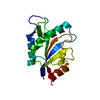
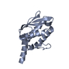
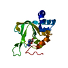
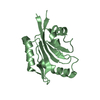
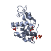
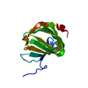
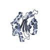
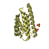
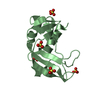
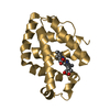
| Title | Structure of Cofilin Mutant (cof1-159p) | ||||||
 Components Components | Cofilin ADF/Cofilin family ADF/Cofilin family | ||||||
 Keywords Keywords | Actin Binding Protein / Actin Depolymerization Factor | ||||||
| Function / homology |  Function and homology information Function and homology informationactin cortical patch / Golgi to plasma membrane protein transport / actin filament severing / actin filament depolymerization / actin filament organization / nuclear matrix / endocytosis / actin filament binding / actin cytoskeleton / plasma membrane / cytoplasmSimilarity search - Function | ||||||
| Biological species |  Saccharomyces cerevisiae (brewer's yeast) Saccharomyces cerevisiae (brewer's yeast) | ||||||
| Method | X-RAY DIFFRACTION / SYNCHROTRON / MOLECULAR REPLACEMENT / Resolution: 1.098 Å | ||||||
 Authors Authors | Kish-Trier, E. / Haarer, B. / Cingolani, G. / Amberg, D.C. | ||||||
 Citation Citation | Journal: To be Published Title: Structure of Cofilin Mutant (cof1-159p) Authors: Kish-Trier, E. / Haarer, B. / Cingolani, G. / Amberg, D.C. | ||||||
| History |
|
- Structure visualization
| Structure viewer | Molecule: MolmilJmol/JSmol |
|---|
- Downloads & links
-Download
| PDBx/mmCIF format | 4kef.cif.gz | 96 KB | Display | PDBx/mmCIF format |
|---|---|---|---|---|
| PDB format | pdb4kef.ent.gz | 74.8 KB | Display | PDB format |
| PDBx/mmJSON format | 4kef.json.gz | Tree view | PDBx/mmJSON format | |
| Others |  Other downloads Other downloads |
-Validation report
| Arichive directory | https://data.pdbj.org/pub/pdb/validation_reports/ke/4kefftp://data.pdbj.org/pub/pdb/validation_reports/ke/4kef | HTTPS FTP |
|---|
-Related structure data
| Related structure data | |
|---|---|
| Similar structure data |


-Links
 PDBj
PDBj
- Assembly
| Deposited unit | 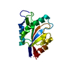
| ||||||||
|---|---|---|---|---|---|---|---|---|---|
| 1 |
| ||||||||
| Unit cell |
|
-Components
| #1: Protein | ADF/Cofilin family / Actin-depolymerizing factor 1 Mass: 15831.506 Da / Num. of mol.: 1 / Mutation: K82D Source method: isolated from a genetically manipulated source Source: (gene. exp.) Saccharomyces cerevisiae (brewer's yeast)Strain: ATCC 204508 / S288c / Gene: COF1, YLL050C, L0596 / Production host:  Escherichia coli (E. coli) / References: UniProt: Q03048 Escherichia coli (E. coli) / References: UniProt: Q03048 |
|---|---|
| #2: Water | ChemComp-HOH / Water Mass: 18.015 Da / Num. of mol.: 184 / Source method: isolated from a natural source / Formula: H2O Mass: 18.015 Da / Num. of mol.: 184 / Source method: isolated from a natural source / Formula: H2O |
-Experimental details
-Experiment
| Experiment | Method: X-RAY DIFFRACTION / Number of used crystals: 1 |
|---|
- Sample preparation
| Crystal | Density Matthews: 1.94 Å3/Da / Density % sol: 36.73 % |
|---|---|
| Crystal grow | Temperature: 293 K / Method: vapor diffusion, hanging drop / pH: 7.4 Details: 20% PEG4000, 150mM NaCl, pH 7.4, VAPOR DIFFUSION, HANGING DROP, temperature 293K |
-Data collection
| Diffraction source | Source: SYNCHROTRON / Site: NSLS  / Beamline: X29A / Wavelength: 1 / Beamline: X29A / Wavelength: 1 |
|---|---|
| Detector | Type: ADSC QUANTUM 315 / Detector: CCD |
| Radiation | Protocol: SINGLE WAVELENGTH / Monochromatic (M) / Laue (L): M / Scattering type: x-ray |
| Radiation wavelength | Wavelength: 1 Å / Relative weight: 1 |
| Reflection | Resolution: 1.098→30 Å / Num. obs: 49213 / % possible obs: 99.77 % / Observed criterion σ(F): 2.8 / Observed criterion σ(I): 2.8 / Redundancy: 4.2 % / Biso Wilson estimate: 10.45 Å2 / Rsym value: 0.076 / Net I/σ(I): 2.8 |
| Reflection shell | Resolution: 1.098→1.14 Å / Redundancy: 2.3 % / Mean I/σ(I) obs: 2.8 / Num. unique all: 205008 / Rsym value: 0.345 / % possible all: 98.99 |
- Processing
| Software |
| |||||||||||||||||||||||||||||||||||||||||||||||||||||||||||||||||||||||||||||||||||||||||||||||||||||||||
|---|---|---|---|---|---|---|---|---|---|---|---|---|---|---|---|---|---|---|---|---|---|---|---|---|---|---|---|---|---|---|---|---|---|---|---|---|---|---|---|---|---|---|---|---|---|---|---|---|---|---|---|---|---|---|---|---|---|---|---|---|---|---|---|---|---|---|---|---|---|---|---|---|---|---|---|---|---|---|---|---|---|---|---|---|---|---|---|---|---|---|---|---|---|---|---|---|---|---|---|---|---|---|---|---|---|---|
| Refinement | Method to determine structure: MOLECULAR REPLACEMENT / Resolution: 1.098→28.697 Å / WRfactor Rfree: 0.1443 / WRfactor Rwork: 0.1265 / SU ML: 0.09 / σ(F): 0 / Phase error: 13.29 / Stereochemistry target values: ML
| |||||||||||||||||||||||||||||||||||||||||||||||||||||||||||||||||||||||||||||||||||||||||||||||||||||||||
| Solvent computation | Shrinkage radii: 0.9 Å / VDW probe radii: 1.11 Å / Solvent model: FLAT BULK SOLVENT MODEL | |||||||||||||||||||||||||||||||||||||||||||||||||||||||||||||||||||||||||||||||||||||||||||||||||||||||||
| Refinement step | Cycle: LAST / Resolution: 1.098→28.697 Å
| |||||||||||||||||||||||||||||||||||||||||||||||||||||||||||||||||||||||||||||||||||||||||||||||||||||||||
| Refine LS restraints |
| |||||||||||||||||||||||||||||||||||||||||||||||||||||||||||||||||||||||||||||||||||||||||||||||||||||||||
| LS refinement shell |
|
