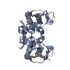
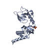
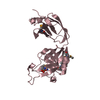
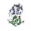
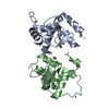
- PDB-4k73: X-ray crystal structure of an L,D-transpeptidase from Mycobacteri... -
+
Open data
ID or keywords:
Loading...
-
Basic information
Entry
Database: PDB / ID: 4k73
Title
X-ray crystal structure of an L,D-transpeptidase from Mycobacterium tuberculosis H37Rv
Components
L,D-transpeptidase
Keywords
TRANSFERASE / Structural Genomics / NIAID / National Institute of Allergy and Infectious Diseases / Seattle Structural Genomics Center for Infectious Disease / SSGCID / L / D-transpeptidase / transpeptidase
Function / homology
Function and homology information
peptidoglycan-protein cross-linking / peptidoglycan L,D-transpeptidase activity / Transferases; Acyltransferases; Aminoacyltransferases / acyltransferase activity / cell wall organization / regulation of cell shape / extracellular region Similarity search - Function
Mass: 18.015 Da / Num. of mol.: 252 / Source method: isolated from a natural source / Formula: H2O
-
Experimental details
-
Experiment
Experiment
Method: X-RAY DIFFRACTION / Number of used crystals: 2
-
Sample preparation
Crystal
Density Matthews: 1.85 Å3/Da / Density % sol: 33.64 %
Crystal grow
Temperature: 289 K / Method: vapor diffusion, sitting drop / pH: 7.5 Details: MCSG1 E9: 0.2 M calcium chloride, 20% PEG3350, pH 7.50, VAPOR DIFFUSION, SITTING DROP, temperature 289K
-
Data collection
Diffraction
ID
Mean temperature (K)
Crystal-ID
1
100
1
2
100
1
Diffraction source
Source
Site
Beamline
Type
ID
Wavelength (Å)
SYNCHROTRON
ALS
5.0.1
1
0.977408
ROTATING ANODE
RIGAKU FR-E+ SUPERBRIGHT
2
1.54
Detector
Type
ID
Detector
Date
ADSC QUANTUM 315
1
CCD
Jan 26, 2013
RIGAKU SATURN 944+
2
CCD
Apr 2, 2013
Radiation
ID
Protocol
Monochromatic (M) / Laue (L)
Scattering type
Wavelength-ID
1
SINGLEWAVELENGTH
M
x-ray
1
2
SINGLEWAVELENGTH
M
x-ray
1
Radiation wavelength
ID
Wavelength (Å)
Relative weight
1
0.977408
1
2
1.54
1
Reflection
Resolution: 1.65→50 Å / Num. all: 28399 / Num. obs: 28136 / % possible obs: 99.1 % / Redundancy: 4.2 % / Rmerge(I) obs: 0.052 / Net I/σ(I): 18.23
-
Processing
Software
Name
Version
Classification
PHENIX
modelbuilding
REFMAC
5.7.0032
refinement
XDS
datareduction
XSCALE
datascaling
PHENIX
phasing
Refinement
Method to determine structure: SAD / Resolution: 1.65→46.48 Å / Cor.coef. Fo:Fc: 0.95 / Cor.coef. Fo:Fc free: 0.932 / SU B: 3.872 / SU ML: 0.073 / Cross valid method: THROUGHOUT / ESU R: 0.106 / ESU R Free: 0.105 / Stereochemistry target values: MAXIMUM LIKELIHOOD / Details: HYDROGENS HAVE BEEN ADDED IN THE RIDING POSITIONS
Rfactor
Num. reflection
% reflection
Selection details
Rfree
0.23077
1416
5 %
RANDOM
Rwork
0.19571
-
-
-
obs
0.19753
26663
99.08 %
-
all
-
28399
-
-
Solvent computation
Ion probe radii: 0.8 Å / Shrinkage radii: 0.8 Å / VDW probe radii: 1.2 Å / Solvent model: MASK
Displacement parameters
Biso mean: 21.014 Å2
Baniso -1
Baniso -2
Baniso -3
1-
-0.95 Å2
-0 Å2
0 Å2
2-
-
-0.35 Å2
0 Å2
3-
-
-
1.31 Å2
Refinement step
Cycle: LAST / Resolution: 1.65→46.48 Å
Protein
Nucleic acid
Ligand
Solvent
Total
Num. atoms
1644
0
1
252
1897
Refine LS restraints
Refine-ID
Type
Dev ideal
Dev ideal target
Number
X-RAY DIFFRACTION
r_bond_refined_d
0.014
0.019
1738
X-RAY DIFFRACTION
r_bond_other_d
0.002
0.02
1576
X-RAY DIFFRACTION
r_angle_refined_deg
1.712
1.94
2391
X-RAY DIFFRACTION
r_angle_other_deg
0.929
3
3624
X-RAY DIFFRACTION
r_dihedral_angle_1_deg
6.3
5
227
X-RAY DIFFRACTION
r_dihedral_angle_2_deg
25.807
23.134
67
X-RAY DIFFRACTION
r_dihedral_angle_3_deg
12.272
15
233
X-RAY DIFFRACTION
r_dihedral_angle_4_deg
16.744
15
9
X-RAY DIFFRACTION
r_chiral_restr
0.093
0.2
271
X-RAY DIFFRACTION
r_gen_planes_refined
0.007
0.021
2006
X-RAY DIFFRACTION
r_gen_planes_other
0.002
0.02
393
X-RAY DIFFRACTION
r_nbd_refined
X-RAY DIFFRACTION
r_nbd_other
X-RAY DIFFRACTION
r_nbtor_refined
X-RAY DIFFRACTION
r_nbtor_other
X-RAY DIFFRACTION
r_xyhbond_nbd_refined
X-RAY DIFFRACTION
r_xyhbond_nbd_other
X-RAY DIFFRACTION
r_metal_ion_refined
X-RAY DIFFRACTION
r_metal_ion_other
X-RAY DIFFRACTION
r_symmetry_vdw_refined
X-RAY DIFFRACTION
r_symmetry_vdw_other
X-RAY DIFFRACTION
r_symmetry_hbond_refined
X-RAY DIFFRACTION
r_symmetry_hbond_other
X-RAY DIFFRACTION
r_symmetry_metal_ion_refined
X-RAY DIFFRACTION
r_symmetry_metal_ion_other
X-RAY DIFFRACTION
r_mcbond_it
0.922
1.362
905
X-RAY DIFFRACTION
r_mcbond_other
0.922
1.361
904
X-RAY DIFFRACTION
r_mcangle_it
1.547
2.035
1133
X-RAY DIFFRACTION
r_scbond_it
1.094
1.467
833
X-RAY DIFFRACTION
r_scangle_it
X-RAY DIFFRACTION
r_rigid_bond_restr
X-RAY DIFFRACTION
r_sphericity_free
X-RAY DIFFRACTION
r_sphericity_bonded
LS refinement shell
Resolution: 1.65→1.693 Å / Total num. of bins used: 20
Rfactor
Num. reflection
% reflection
Rfree
0.288
106
-
Rwork
0.269
1950
-
obs
-
-
99.61 %
Refinement TLS params.
Method: refined / Origin x: -9.2092 Å / Origin y: 2.3378 Å / Origin z: -10.9587 Å
11
12
13
21
22
23
31
32
33
T
0.0438 Å2
-0.0146 Å2
0.0106 Å2
-
0.0374 Å2
-0.0139 Å2
-
-
0.0414 Å2
L
1.6972 °2
-0.1221 °2
0.3693 °2
-
0.1601 °2
-0.103 °2
-
-
0.5965 °2
S
-0.0244 Å °
0.2222 Å °
-0.0422 Å °
-0.0641 Å °
0.0159 Å °
-0.0377 Å °
-0.0092 Å °
0.0023 Å °
0.0085 Å °
+
About Yorodumi
-
News
-
Feb 9, 2022. New format data for meta-information of EMDB entries
New format data for meta-information of EMDB entries
Version 3 of the EMDB header file is now the official format.
The previous official version 1.9 will be removed from the archive.
In the structure databanks used in Yorodumi, some data are registered as the other names, "COVID-19 virus" and "2019-nCoV". Here are the details of the virus and the list of structure data.
Jan 31, 2019. EMDB accession codes are about to change! (news from PDBe EMDB page)
EMDB accession codes are about to change! (news from PDBe EMDB page)
The allocation of 4 digits for EMDB accession codes will soon come to an end. Whilst these codes will remain in use, new EMDB accession codes will include an additional digit and will expand incrementally as the available range of codes is exhausted. The current 4-digit format prefixed with “EMD-” (i.e. EMD-XXXX) will advance to a 5-digit format (i.e. EMD-XXXXX), and so on. It is currently estimated that the 4-digit codes will be depleted around Spring 2019, at which point the 5-digit format will come into force.
The EM Navigator/Yorodumi systems omit the EMD- prefix.
Related info.:Q: What is EMD? / ID/Accession-code notation in Yorodumi/EM Navigator
Yorodumi is a browser for structure data from EMDB, PDB, SASBDB, etc.
This page is also the successor to EM Navigator detail page, and also detail information page/front-end page for Omokage search.
The word "yorodu" (or yorozu) is an old Japanese word meaning "ten thousand". "mi" (miru) is to see.
Related info.:EMDB / PDB / SASBDB / Comparison of 3 databanks / Yorodumi Search / Aug 31, 2016. New EM Navigator & Yorodumi / Yorodumi Papers / Jmol/JSmol / Function and homology information / Changes in new EM Navigator and Yorodumi
 Movie
Movie Controller

 Yorodumi
 Open data
 Basic information
 Components
 Keywords
 TRANSFERASE /
  Function and homology information

 Authors
 Citation
 Structure visualization
 Downloads & links
 Other downloads
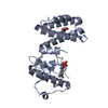


 PDBj
PDBj Assembly
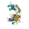

 Mass: 40.078 Da / Num. of mol.: 1 / Source method: obtained synthetically / Formula: Ca
 Mass: 18.015 Da / Num. of mol.: 252 / Source method: isolated from a natural source / Formula: H2O
 Sample preparation
 Processing