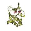
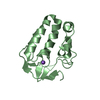
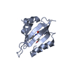
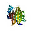
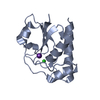
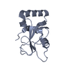
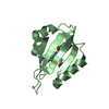
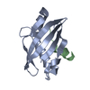
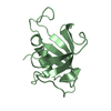
PKG II's Amino Terminal Cyclic Nucleotide Binding Domain (CNB-A) in a complex with cAMP
Components
cGMP-dependent protein kinase 2
Keywords
PROTEIN BINDING / Binding Sites / Cyclic AMP / Cyclic GMP / Cyclic GMP-Dependent Protein Kinase Type II / Mutagenesis / Site-Directed
Function / homology
Function and homology information
negative regulation of chloride transport / tetrahydrobiopterin metabolic process / cGMP-dependent protein kinase / cGMP-dependent protein kinase activity / positive regulation of chondrocyte differentiation / mitogen-activated protein kinase binding / positive regulation of protein localization / cGMP effects / cGMP binding / Tetrahydrobiopterin (BH4) synthesis, recycling, salvage and regulation ...negative regulation of chloride transport / tetrahydrobiopterin metabolic process / cGMP-dependent protein kinase / cGMP-dependent protein kinase activity / positive regulation of chondrocyte differentiation / mitogen-activated protein kinase binding / positive regulation of protein localization / cGMP effects / cGMP binding / Tetrahydrobiopterin (BH4) synthesis, recycling, salvage and regulation / protein localization to plasma membrane / RAS processing / Ca2+ pathway / nuclear membrane / protein kinase activity / apical plasma membrane / protein phosphorylation / protein serine kinase activity / signal transduction / ATP binding / identical protein binding / cytosol Similarity search - Function
cGMP-dependent kinase / cGMP-dependent protein kinase, catalytic domain / Cyclic nucleotide-binding domain signature 2. / Cyclic nucleotide-binding domain signature 1. / Cyclic nucleotide-binding, conserved site / Cyclic nucleotide-monophosphate binding domain / Cyclic nucleotide-binding domain / cAMP/cGMP binding motif profile. / Cyclic nucleotide-binding domain / Cyclic nucleotide-binding domain superfamily ...cGMP-dependent kinase / cGMP-dependent protein kinase, catalytic domain / Cyclic nucleotide-binding domain signature 2. / Cyclic nucleotide-binding domain signature 1. / Cyclic nucleotide-binding, conserved site / Cyclic nucleotide-monophosphate binding domain / Cyclic nucleotide-binding domain / cAMP/cGMP binding motif profile. / Cyclic nucleotide-binding domain / Cyclic nucleotide-binding domain superfamily / Extension to Ser/Thr-type protein kinases / Jelly Rolls / AGC-kinase, C-terminal / AGC-kinase C-terminal domain profile. / RmlC-like jelly roll fold / Jelly Rolls / Serine/threonine-protein kinase, active site / Serine/Threonine protein kinases active-site signature. / Protein kinase domain / Serine/Threonine protein kinases, catalytic domain / Protein kinase, ATP binding site / Protein kinases ATP-binding region signature. / Protein kinase domain profile. / Protein kinase domain / Protein kinase-like domain superfamily / Sandwich / Mainly Beta Similarity search - Domain/homology
Mass: 18.015 Da / Num. of mol.: 106 / Source method: isolated from a natural source / Formula: H2O
-
Experimental details
-
Experiment
Experiment
Method: X-RAY DIFFRACTION / Number of used crystals: 1
-
Sample preparation
Crystal
Density Matthews: 2.24 Å3/Da / Density % sol: 44.98 %
Crystal grow
Temperature: 296 K / Method: vapor diffusion, hanging drop Details: 200 mM calcium chloride, 200 mM cadmium chloride, 200 mM cobalt (II) chloride, 20% polyethylene glycol 3350
In the structure databanks used in Yorodumi, some data are registered as the other names, "COVID-19 virus" and "2019-nCoV". Here are the details of the virus and the list of structure data.
Jan 31, 2019. EMDB accession codes are about to change! (news from PDBe EMDB page)
EMDB accession codes are about to change! (news from PDBe EMDB page)
The allocation of 4 digits for EMDB accession codes will soon come to an end. Whilst these codes will remain in use, new EMDB accession codes will include an additional digit and will expand incrementally as the available range of codes is exhausted. The current 4-digit format prefixed with “EMD-” (i.e. EMD-XXXX) will advance to a 5-digit format (i.e. EMD-XXXXX), and so on. It is currently estimated that the 4-digit codes will be depleted around Spring 2019, at which point the 5-digit format will come into force.
The EM Navigator/Yorodumi systems omit the EMD- prefix.
Related info.:Q: What is EMD? / ID/Accession-code notation in Yorodumi/EM Navigator
Yorodumi is a browser for structure data from EMDB, PDB, SASBDB, etc.
This page is also the successor to EM Navigator detail page, and also detail information page/front-end page for Omokage search.
The word "yorodu" (or yorozu) is an old Japanese word meaning "ten thousand". "mi" (miru) is to see.
Related info.:EMDB / PDB / SASBDB / Comparison of 3 databanks / Yorodumi Search / Aug 31, 2016. New EM Navigator & Yorodumi / Yorodumi Papers / Jmol/JSmol / Function and homology information / Changes in new EM Navigator and Yorodumi
 Movie
Movie Controller

 Yorodumi
 Open data
 Basic information
 Components
 Keywords
 PROTEIN BINDING /
  Function and homology information
 Authors
 United States, 1items
  Citation
 Structure visualization
 Downloads & links
 Other downloads
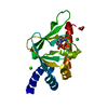


 PDBj
PDBj


 Assembly

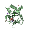


 Mass: 112.411 Da / Num. of mol.: 6 / Source method: obtained synthetically / Formula: Cd
 Mass: 329.206 Da / Num. of mol.: 2 / Source method: obtained synthetically / Formula: C10H12N5O6P
 Mass: 58.933 Da / Num. of mol.: 4 / Source method: obtained synthetically / Formula: Co
 Mass: 40.078 Da / Num. of mol.: 2 / Source method: isolated from a natural source / Formula: Ca
 Mass: 62.068 Da / Num. of mol.: 1 / Source method: obtained synthetically / Formula: C2H6O2
 Mass: 22.990 Da / Num. of mol.: 1 / Source method: isolated from a natural source / Formula: Na
 Sample preparation
 Processing