 Movie
Movie Controller


+ Open data

- Basic information
| Entry | Database: PDB / ID: 4k3b | ||||||
|---|---|---|---|---|---|---|---|
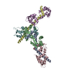
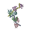
| Title | The crystal structure of BamA from Neisseria gonorrhoeae | ||||||
 Components Components | Outer membrane protein assembly factor BamA | ||||||
 Keywords Keywords |  MEMBRANE PROTEIN / beta-barrel membrane protein / insertase MEMBRANE PROTEIN / beta-barrel membrane protein / insertase | ||||||
| Function / homology |  Function and homology information Function and homology informationGram-negative-bacterium-type cell outer membrane assembly / protein insertion into membrane / cell outer membrane / membrane => GO:0016020 Similarity search - Function | ||||||
| Biological species |  Neisseria gonorrhoeae (bacteria) Neisseria gonorrhoeae (bacteria) | ||||||
| Method | X-RAY DIFFRACTION / SYNCHROTRON / MOLECULAR REPLACEMENT / Resolution: 3.2 Å | ||||||
 Authors Authors | Noinaj, N. / Lukacik, P. / Chang, H. / Easley, N. / Buchanan, S.K. | ||||||
 Citation Citation | Journal: Nature / Year: 2013 Title: Structural insight into the biogenesis of beta-barrel membrane proteins. Authors: Noinaj, N. / Kuszak, A.J. / Gumbart, J.C. / Lukacik, P. / Chang, H. / Easley, N.C. / Lithgow, T. / Buchanan, S.K. | ||||||
| History |
|
- Structure visualization
| Structure viewer | Molecule: MolmilJmol/JSmol |
|---|
- Downloads & links
-Download
| PDBx/mmCIF format | 4k3b.cif.gz | 306 KB | Display | PDBx/mmCIF format |
|---|---|---|---|---|
| PDB format | pdb4k3b.ent.gz | 251.3 KB | Display | PDB format |
| PDBx/mmJSON format | 4k3b.json.gz | Tree view | PDBx/mmJSON format | |
| Others |  Other downloads Other downloads |
-Validation report
| Arichive directory | https://data.pdbj.org/pub/pdb/validation_reports/k3/4k3bftp://data.pdbj.org/pub/pdb/validation_reports/k3/4k3b | HTTPS FTP |
|---|
-Related structure data


-Links
 PDBj
PDBj- Assembly
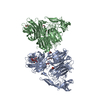
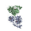
| Deposited unit | 
| ||||||||
|---|---|---|---|---|---|---|---|---|---|
| 1 |
| ||||||||
| Unit cell |
|
-Components
| #1: Protein | Mass: 88047.078 Da / Num. of mol.: 1 Source method: isolated from a genetically manipulated source Source: (gene. exp.) Neisseria gonorrhoeae (bacteria) / Strain: FA 1090 / Gene: bamA, NGO1801 / Production host: Escherichia coli (E. coli) / References: UniProt: Q5F5W8 |
|---|
-Experimental details
-Experiment
| Experiment | Method: X-RAY DIFFRACTION / Number of used crystals: 1 |
|---|
- Sample preparation
| Crystal | Density Matthews: 4.35 Å3/Da / Density % sol: 71.74 % |
|---|---|
| Crystal grow | Temperature: 295 K / pH: 7.5 Details: 0.1 M NaCl, 0.1 M K-phosphate 7.0, 32% PEG 300, and 200 mM Na-malonate, pH 7.5, temperature 295K |
-Data collection
| Diffraction | Mean temperature: 100 K |
|---|---|
| Diffraction source | Source: SYNCHROTRON / Site: APS  / Beamline: 22-ID / Wavelength: 1 Å / Beamline: 22-ID / Wavelength: 1 Å |
| Detector | Type: MARMOSAIC 300 mm CCD / Detector: CCD / Date: Jul 1, 2011 |
| Radiation | Monochromator: double crystal / Protocol: SINGLE WAVELENGTH / Monochromatic (M) / Laue (L): M / Scattering type: x-ray |
| Radiation wavelength | Wavelength: 1 Å / Relative weight: 1 |
| Reflection | Resolution: 3.2→50 Å / Num. all: 26110 / Num. obs: 25674 / % possible obs: 99.5 % / Observed criterion σ(F): 1 / Observed criterion σ(I): 1 / Redundancy: 6.2 % / Rsym value: 0.14 / Net I/σ(I): 17.2 |
| Reflection shell | Resolution: 3.2→3.31 Å / Redundancy: 5.4 % / Mean I/σ(I) obs: 1.8 / Num. unique all: 2582 / Rsym value: 0.957 / % possible all: 99.2 |
- Processing
| Software |
| |||||||||||||||||||||||||||||||||||||||||||||||||||||||||||||||||||||||||||||||||||||||||||||||||||||||||
|---|---|---|---|---|---|---|---|---|---|---|---|---|---|---|---|---|---|---|---|---|---|---|---|---|---|---|---|---|---|---|---|---|---|---|---|---|---|---|---|---|---|---|---|---|---|---|---|---|---|---|---|---|---|---|---|---|---|---|---|---|---|---|---|---|---|---|---|---|---|---|---|---|---|---|---|---|---|---|---|---|---|---|---|---|---|---|---|---|---|---|---|---|---|---|---|---|---|---|---|---|---|---|---|---|---|---|
| Refinement | Method to determine structure: MOLECULAR REPLACEMENT / Resolution: 3.2→14.999 Å / SU ML: 0.53 / σ(F): 1.35 / Phase error: 30.41 / Stereochemistry target values: ML
| |||||||||||||||||||||||||||||||||||||||||||||||||||||||||||||||||||||||||||||||||||||||||||||||||||||||||
| Solvent computation | Shrinkage radii: 0.9 Å / VDW probe radii: 1.11 Å / Solvent model: FLAT BULK SOLVENT MODEL | |||||||||||||||||||||||||||||||||||||||||||||||||||||||||||||||||||||||||||||||||||||||||||||||||||||||||
| Refinement step | Cycle: LAST / Resolution: 3.2→14.999 Å
| |||||||||||||||||||||||||||||||||||||||||||||||||||||||||||||||||||||||||||||||||||||||||||||||||||||||||
| Refine LS restraints |
| |||||||||||||||||||||||||||||||||||||||||||||||||||||||||||||||||||||||||||||||||||||||||||||||||||||||||
| LS refinement shell |
| |||||||||||||||||||||||||||||||||||||||||||||||||||||||||||||||||||||||||||||||||||||||||||||||||||||||||
| Refinement TLS params. | Method: refined / Refine-ID: X-RAY DIFFRACTION
| |||||||||||||||||||||||||||||||||||||||||||||||||||||||||||||||||||||||||||||||||||||||||||||||||||||||||
| Refinement TLS group |
|
