 Movie
Movie Controller


+ Open data

- Basic information
| Entry | Database: PDB / ID: 4j8m | ||||||
|---|---|---|---|---|---|---|---|
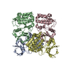
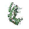
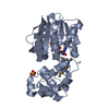
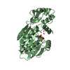
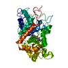
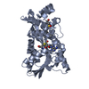
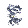
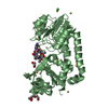
| Title | Aurora A in complex with CD532 | ||||||
 Components Components | Aurora kinase A | ||||||
 Keywords Keywords | TRANSFERASE/TRANSFERASE INHIBITOR / Aurora A / type II / kinase inhibitor / TRANSFERASE-TRANSFERASE INHIBITOR complex | ||||||
| Function / homology |  Function and homology information Function and homology informationInteraction between PHLDA1 and AURKA / regulation of centrosome cycle / axon hillock / spindle assembly involved in female meiosis I / cilium disassembly / spindle pole centrosome / positive regulation of oocyte maturation / histone H3S10 kinase activity / chromosome passenger complex / pronucleus ...Interaction between PHLDA1 and AURKA / regulation of centrosome cycle / axon hillock / spindle assembly involved in female meiosis I / cilium disassembly / spindle pole centrosome / positive regulation of oocyte maturation / histone H3S10 kinase activity / chromosome passenger complex / pronucleus / meiotic spindle / mitotic centrosome separation / germinal vesicle / protein localization to centrosome / anterior/posterior axis specification / centrosome localization / neuron projection extension / positive regulation of mitochondrial fission / spindle organization / mitotic spindle pole / SUMOylation of DNA replication proteins / spindle midzone / regulation of G2/M transition of mitotic cell cycle / centriole / protein serine/threonine/tyrosine kinase activity / positive regulation of mitotic cell cycle / AURKA Activation by TPX2 / mitotic spindle organization / positive regulation of mitotic nuclear division / TP53 Regulates Transcription of Genes Involved in G2 Cell Cycle Arrest / ciliary basal body / negative regulation of protein binding / regulation of cytokinesis / regulation of signal transduction by p53 class mediator / molecular function activator activity / liver regeneration / FBXL7 down-regulates AURKA during mitotic entry and in early mitosis / regulation of protein stability / APC/C:Cdh1 mediated degradation of Cdc20 and other APC/C:Cdh1 targeted proteins in late mitosis/early G1 / mitotic spindle / kinetochore / spindle / response to wounding / microtubule cytoskeleton / G2/M transition of mitotic cell cycle / Regulation of PLK1 Activity at G2/M Transition / positive regulation of proteasomal ubiquitin-dependent protein catabolic process / mitotic cell cycle / midbody / proteasome-mediated ubiquitin-dependent protein catabolic process / basolateral plasma membrane / peptidyl-serine phosphorylation / Regulation of TP53 Activity through Phosphorylation / microtubule / postsynaptic density / protein autophosphorylation / non-specific serine/threonine protein kinase / protein kinase activity / protein heterodimerization activity / cell division / protein phosphorylation / negative regulation of gene expression / protein serine kinase activity / protein serine/threonine kinase activity / centrosome / glutamatergic synapse / apoptotic process / ubiquitin protein ligase binding / negative regulation of apoptotic process / protein kinase binding / perinuclear region of cytoplasm / nucleoplasm / ATP binding / nucleus / cytosolSimilarity search - Function | ||||||
| Biological species |  Homo sapiens (human) Homo sapiens (human) | ||||||
| Method | X-RAY DIFFRACTION / SYNCHROTRON / MOLECULAR REPLACEMENT / molecular replacement / Resolution: 1.853 Å | ||||||
 Authors Authors | Meyerowitz, J.G. / Gustafson, W.C. / Shokat, K.M. / Weiss, W.A. | ||||||
 Citation Citation | Journal: Cancer Cell / Year: 2014 Title: Drugging MYCN through an Allosteric Transition in Aurora Kinase A. Authors: Gustafson, W.C. / Meyerowitz, J.G. / Nekritz, E.A. / Chen, J. / Benes, C. / Charron, E. / Simonds, E.F. / Seeger, R. / Matthay, K.K. / Hertz, N.T. / Eilers, M. / Shokat, K.M. / Weiss, W.A. | ||||||
| History |
|
- Structure visualization
| Structure viewer | Molecule: MolmilJmol/JSmol |
|---|
- Downloads & links
-Download
| PDBx/mmCIF format | 4j8m.cif.gz | 174.5 KB | Display | PDBx/mmCIF format |
|---|---|---|---|---|
| PDB format | pdb4j8m.ent.gz | 148.4 KB | Display | PDB format |
| PDBx/mmJSON format | 4j8m.json.gz | Tree view | PDBx/mmJSON format | |
| Others |  Other downloads Other downloads |
-Validation report
| Arichive directory | https://data.pdbj.org/pub/pdb/validation_reports/j8/4j8mftp://data.pdbj.org/pub/pdb/validation_reports/j8/4j8m | HTTPS FTP |
|---|
-Related structure data


-Links
 PDBj
PDBj


- Assembly
| Deposited unit | 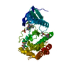
| |||||||||
|---|---|---|---|---|---|---|---|---|---|---|
| 1 |
| |||||||||
| Unit cell |
| |||||||||
| Components on special symmetry positions |
|
-Components
-Protein , 1 types, 1 molecules A
| #1: Protein | / Aurora 2 / Aurora/IPL1-related kinase 1 / ARK-1 / Aurora-related kinase 1 / hARK1 / Breast tumor- ...Aurora 2 / Aurora/IPL1-related kinase 1 / ARK-1 / Aurora-related kinase 1 / hARK1 / Breast tumor-amplified kinase / Serine/threonine-protein kinase 15 / Serine/threonine-protein kinase 6 / Serine/threonine-protein kinase aurora-A Mass: 32479.104 Da / Num. of mol.: 1 Source method: isolated from a genetically manipulated source Source: (gene. exp.) Homo sapiens (human)Gene: AIK, AIRK1, ARK1, AURA, AURKA, AYK1, BTAK, IAK1, STK15, STK6 Plasmid: pET28b / Production host:  Escherichia coli (E. coli) / Strain (production host): BL21(DE3) Escherichia coli (E. coli) / Strain (production host): BL21(DE3)References: UniProt: O14965, non-specific serine/threonine protein kinase |
|---|
-Non-polymers , 6 types, 178 molecules 


| #2: Chemical | ChemComp-CJ5 /  Mass: 522.525 Da / Num. of mol.: 1 / Source method: obtained synthetically / Formula: C26H25F3N8O Mass: 522.525 Da / Num. of mol.: 1 / Source method: obtained synthetically / Formula: C26H25F3N8O | ||||
|---|---|---|---|---|---|
| #3: Chemical | ChemComp-MG /  Mass: 24.305 Da / Num. of mol.: 1 / Source method: obtained synthetically / Formula: Mg Mass: 24.305 Da / Num. of mol.: 1 / Source method: obtained synthetically / Formula: Mg | ||||
| #4: Chemical | ChemComp-PO4 / Phosphate Mass: 94.971 Da / Num. of mol.: 1 / Source method: obtained synthetically / Formula: PO4 Mass: 94.971 Da / Num. of mol.: 1 / Source method: obtained synthetically / Formula: PO4 | ||||
| #5: Chemical | Dimethyl sulfoxide Mass: 78.133 Da / Num. of mol.: 2 / Source method: obtained synthetically / Formula: C2H6OS / Comment: DMSO, precipitant*YM Mass: 78.133 Da / Num. of mol.: 2 / Source method: obtained synthetically / Formula: C2H6OS / Comment: DMSO, precipitant*YM#6: Chemical | ChemComp-EDO / | Ethylene glycol Mass: 62.068 Da / Num. of mol.: 1 / Source method: obtained synthetically / Formula: C2H6O2 Mass: 62.068 Da / Num. of mol.: 1 / Source method: obtained synthetically / Formula: C2H6O2#7: Water | ChemComp-HOH / | WaterMass: 18.015 Da / Num. of mol.: 172 / Source method: isolated from a natural source / Formula: H2O |
-Experimental details
-Experiment
| Experiment | Method: X-RAY DIFFRACTION / Number of used crystals: 1 |
|---|
- Sample preparation
| Crystal | Density Matthews: 2.22 Å3/Da / Density % sol: 44.51 % |
|---|---|
| Crystal grow | Temperature: 298 K / Method: vapor diffusion, hanging drop / pH: 6 Details: 20 mg/ml Aurora A protein, 1 mM CD532, 20% (w/v) PEG8000, 0.2 M magnesium acetate tetrahydrate, 0.1 M sodium cacodylate trihydrate, pH 6.0, VAPOR DIFFUSION, HANGING DROP, temperature 298K |
-Data collection
| Diffraction | Mean temperature: 100 K | |||||||||||||||||||||||||||||||||||||||||||||||||||||||||||||||||||||||||||||||||||||||||||||||||||||||||
|---|---|---|---|---|---|---|---|---|---|---|---|---|---|---|---|---|---|---|---|---|---|---|---|---|---|---|---|---|---|---|---|---|---|---|---|---|---|---|---|---|---|---|---|---|---|---|---|---|---|---|---|---|---|---|---|---|---|---|---|---|---|---|---|---|---|---|---|---|---|---|---|---|---|---|---|---|---|---|---|---|---|---|---|---|---|---|---|---|---|---|---|---|---|---|---|---|---|---|---|---|---|---|---|---|---|---|
| Diffraction source | Source: SYNCHROTRON / Site: ALS  / Beamline: 8.2.2 / Wavelength: 1.0088 Å / Beamline: 8.2.2 / Wavelength: 1.0088 Å | |||||||||||||||||||||||||||||||||||||||||||||||||||||||||||||||||||||||||||||||||||||||||||||||||||||||||
| Detector | Type: ADSC QUANTUM 315r / Detector: CCD / Date: Aug 1, 2012 | |||||||||||||||||||||||||||||||||||||||||||||||||||||||||||||||||||||||||||||||||||||||||||||||||||||||||
| Radiation | Monochromator: Double crystal, Si(111) / Protocol: SINGLE WAVELENGTH / Monochromatic (M) / Laue (L): M / Scattering type: x-ray | |||||||||||||||||||||||||||||||||||||||||||||||||||||||||||||||||||||||||||||||||||||||||||||||||||||||||
| Radiation wavelength | Wavelength: 1.0088 Å / Relative weight: 1 | |||||||||||||||||||||||||||||||||||||||||||||||||||||||||||||||||||||||||||||||||||||||||||||||||||||||||
| Reflection | Resolution: 1.85→30 Å / Num. all: 24605 / Num. obs: 24605 / % possible obs: 99 % / Observed criterion σ(F): 0 / Observed criterion σ(I): -3 / Redundancy: 6.5 % / Rmerge(I) obs: 0.106 / Net I/σ(I): 11.6 | |||||||||||||||||||||||||||||||||||||||||||||||||||||||||||||||||||||||||||||||||||||||||||||||||||||||||
| Reflection shell |
|
-Phasing
| Phasing | Method: molecular replacement | |||||||||
|---|---|---|---|---|---|---|---|---|---|---|
| Phasing MR |
|
- Processing
| Software |
| ||||||||||||||||||||||||||||||||||||||||||||||||||||||||||||||||||||||||||||||||||||||||||||||||||||||||||||||||||||||||||||||||||||||||||||||||||||||||||||||||||||||||||||||||||||||||||||||||||||||||
|---|---|---|---|---|---|---|---|---|---|---|---|---|---|---|---|---|---|---|---|---|---|---|---|---|---|---|---|---|---|---|---|---|---|---|---|---|---|---|---|---|---|---|---|---|---|---|---|---|---|---|---|---|---|---|---|---|---|---|---|---|---|---|---|---|---|---|---|---|---|---|---|---|---|---|---|---|---|---|---|---|---|---|---|---|---|---|---|---|---|---|---|---|---|---|---|---|---|---|---|---|---|---|---|---|---|---|---|---|---|---|---|---|---|---|---|---|---|---|---|---|---|---|---|---|---|---|---|---|---|---|---|---|---|---|---|---|---|---|---|---|---|---|---|---|---|---|---|---|---|---|---|---|---|---|---|---|---|---|---|---|---|---|---|---|---|---|---|---|---|---|---|---|---|---|---|---|---|---|---|---|---|---|---|---|---|---|---|---|---|---|---|---|---|---|---|---|---|---|---|---|---|
| Refinement | Method to determine structure: MOLECULAR REPLACEMENT / Resolution: 1.853→29.075 Å / Occupancy max: 1 / Occupancy min: 0 / SU ML: 0.18 / σ(F): 1.42 / Phase error: 20.39 / Stereochemistry target values: ML
| ||||||||||||||||||||||||||||||||||||||||||||||||||||||||||||||||||||||||||||||||||||||||||||||||||||||||||||||||||||||||||||||||||||||||||||||||||||||||||||||||||||||||||||||||||||||||||||||||||||||||
| Solvent computation | Shrinkage radii: 0.9 Å / VDW probe radii: 1.1 Å / Solvent model: FLAT BULK SOLVENT MODEL | ||||||||||||||||||||||||||||||||||||||||||||||||||||||||||||||||||||||||||||||||||||||||||||||||||||||||||||||||||||||||||||||||||||||||||||||||||||||||||||||||||||||||||||||||||||||||||||||||||||||||
| Displacement parameters | Biso mean: 42.2172 Å2 | ||||||||||||||||||||||||||||||||||||||||||||||||||||||||||||||||||||||||||||||||||||||||||||||||||||||||||||||||||||||||||||||||||||||||||||||||||||||||||||||||||||||||||||||||||||||||||||||||||||||||
| Refinement step | Cycle: LAST / Resolution: 1.853→29.075 Å
| ||||||||||||||||||||||||||||||||||||||||||||||||||||||||||||||||||||||||||||||||||||||||||||||||||||||||||||||||||||||||||||||||||||||||||||||||||||||||||||||||||||||||||||||||||||||||||||||||||||||||
| Refine LS restraints |
| ||||||||||||||||||||||||||||||||||||||||||||||||||||||||||||||||||||||||||||||||||||||||||||||||||||||||||||||||||||||||||||||||||||||||||||||||||||||||||||||||||||||||||||||||||||||||||||||||||||||||
| LS refinement shell |
| ||||||||||||||||||||||||||||||||||||||||||||||||||||||||||||||||||||||||||||||||||||||||||||||||||||||||||||||||||||||||||||||||||||||||||||||||||||||||||||||||||||||||||||||||||||||||||||||||||||||||
| Refinement TLS params. | Method: refined / Refine-ID: X-RAY DIFFRACTION
| ||||||||||||||||||||||||||||||||||||||||||||||||||||||||||||||||||||||||||||||||||||||||||||||||||||||||||||||||||||||||||||||||||||||||||||||||||||||||||||||||||||||||||||||||||||||||||||||||||||||||
| Refinement TLS group |
|
