 Movie
Movie Controller

[English] 日本語
 Yorodumi
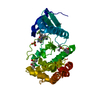
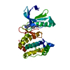
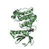
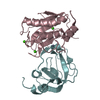
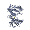
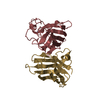
- PDB-4jmf: Crystal structure of ExoT (residues 28 -77)- SpcS complex from Ps... -
+ Open data

- Basic information
| Entry | Database: PDB / ID: 4jmf | ||||||
|---|---|---|---|---|---|---|---|
| Title | Crystal structure of ExoT (residues 28 -77)- SpcS complex from Pseudomonas aeruginosa at 2.1 angstrom | ||||||
 Components Components |
| ||||||
 Keywords Keywords | TOXIN/CHAPERONE /  Type III secretion system / T3SS / virulent effector / TOXIN-CHAPERONE complex Type III secretion system / T3SS / virulent effector / TOXIN-CHAPERONE complex | ||||||
| Function / homology |  Function and homology information Function and homology informationNAD+-protein-arginine ADP-ribosyltransferase / NAD+-protein-arginine ADP-ribosyltransferase activity / protein secretion by the type III secretion system / NAD+ ADP-ribosyltransferase activity / nucleotidyltransferase activity / GTPase activator activity / toxin activity / extracellular regionSimilarity search - Function | ||||||
| Biological species |   Pseudomonas aeruginosa (bacteria) Pseudomonas aeruginosa (bacteria) | ||||||
| Method | X-RAY DIFFRACTION / MIR / Resolution: 2.099 Å | ||||||
 Authors Authors | Datta, S. / Dey, S. | ||||||
 Citation Citation | Journal: Febs J. / Year: 2014 Title: Interfacial residues of SpcS chaperone affects binding of effector toxin ExoT in Pseudomonas aeruginosa: novel insights from structural and computational studies Authors: Dey, S. / Datta, S. | ||||||
| History |
| ||||||
| Remark 650 | HELIX Determination method: Author determined | ||||||
| Remark 700 | SHEET Determination method: Author determined |
- Structure visualization
| Structure viewer | Molecule: MolmilJmol/JSmol |
|---|
- Downloads & links
-Download
| PDBx/mmCIF format | 4jmf.cif.gz | 71.1 KB | Display | PDBx/mmCIF format |
|---|---|---|---|---|
| PDB format | pdb4jmf.ent.gz | 53.3 KB | Display | PDB format |
| PDBx/mmJSON format | 4jmf.json.gz | Tree view | PDBx/mmJSON format | |
| Others |  Other downloads Other downloads |
-Validation report
| Arichive directory | https://data.pdbj.org/pub/pdb/validation_reports/jm/4jmfftp://data.pdbj.org/pub/pdb/validation_reports/jm/4jmf | HTTPS FTP |
|---|
-Related structure data
| Similar structure data |
|---|
-Links
 PDBj
PDBj- Assembly
| Deposited unit | 
| |||||||||
|---|---|---|---|---|---|---|---|---|---|---|
| 1 |
| |||||||||
| Unit cell |
| |||||||||
| Components on special symmetry positions |
|
-Components
| #1: Protein/peptide | Mass: 5416.263 Da / Num. of mol.: 1 / Fragment: UNP residues 28-77 Source method: isolated from a genetically manipulated source Source: (gene. exp.) Pseudomonas aeruginosa (bacteria) / Strain: PA01 / Gene: exoT / Plasmid: pETDuet-1 / Production host: Escherichia coli (E. coli) / Strain (production host): BL21(DE3) / References: UniProt: Q9I788 | ||||
|---|---|---|---|---|---|
| #2: Protein | Mass: 13171.843 Da / Num. of mol.: 2 Source method: isolated from a genetically manipulated source Source: (gene. exp.) Pseudomonas aeruginosa (bacteria) / Strain: PA01 / Gene: SpcS / Plasmid: pETDuet-1 / Production host: Escherichia coli (E. coli) / Strain (production host): BL21(DE3) / References: UniProt: G3XD93#3: Chemical | ChemComp-GOL / | Glycerol  Mass: 92.094 Da / Num. of mol.: 1 / Source method: obtained synthetically / Formula: C3H8O3 Mass: 92.094 Da / Num. of mol.: 1 / Source method: obtained synthetically / Formula: C3H8O3#4: Water | ChemComp-HOH / | Water Mass: 18.015 Da / Num. of mol.: 134 / Source method: isolated from a natural source / Formula: H2O Mass: 18.015 Da / Num. of mol.: 134 / Source method: isolated from a natural source / Formula: H2O |
-Experimental details
-Experiment
| Experiment | Method: X-RAY DIFFRACTION / Number of used crystals: 1 |
|---|
- Sample preparation
| Crystal | Density Matthews: 2.71 Å3/Da / Density % sol: 54.57 % |
|---|---|
| Crystal grow | Temperature: 295 K / Method: vapor diffusion / pH: 6.5 Details: 28% PEGMME 5000, 0.2M ammonium sulphate, pH 6.5, vapour diffusion, temperature 295K, VAPOR DIFFUSION |
-Data collection
| Diffraction | Mean temperature: 100 K | ||||||||||||||||||
|---|---|---|---|---|---|---|---|---|---|---|---|---|---|---|---|---|---|---|---|
| Diffraction source | Source: ROTATING ANODE / Type: RIGAKU ULTRAX 18 / Wavelength: 1.5418 Å | ||||||||||||||||||
| Detector | Type: RIGAKU RAXIS IV++ / Detector: IMAGE PLATE / Date: Aug 5, 2012 | ||||||||||||||||||
| Radiation | Monochromator: Ni Filter / Protocol: SINGLE WAVELENGTH / Monochromatic (M) / Laue (L): M / Scattering type: x-ray | ||||||||||||||||||
| Radiation wavelength | Wavelength: 1.5418 Å / Relative weight: 1 | ||||||||||||||||||
| Reflection | Resolution: 2.099→40 Å / Num. all: 21477 / Num. obs: 21244 / % possible obs: 99 % / Observed criterion σ(F): 1 / Observed criterion σ(I): 1 / Biso Wilson estimate: 34.09 Å2 | ||||||||||||||||||
| Reflection shell |
|
- Processing
| Software |
| |||||||||||||||||||||||||||||||||||||||||||||||||||||||||||||||||||||||||||||||||||||||||||||||||||||||||
|---|---|---|---|---|---|---|---|---|---|---|---|---|---|---|---|---|---|---|---|---|---|---|---|---|---|---|---|---|---|---|---|---|---|---|---|---|---|---|---|---|---|---|---|---|---|---|---|---|---|---|---|---|---|---|---|---|---|---|---|---|---|---|---|---|---|---|---|---|---|---|---|---|---|---|---|---|---|---|---|---|---|---|---|---|---|---|---|---|---|---|---|---|---|---|---|---|---|---|---|---|---|---|---|---|---|---|
| Refinement | Method to determine structure: MIR / Resolution: 2.099→30.482 Å / Occupancy max: 1 / Occupancy min: 1 / FOM work R set: 0.8389 / SU ML: 0.21 / σ(F): 0 / Phase error: 22.38 / Stereochemistry target values: MLHL
| |||||||||||||||||||||||||||||||||||||||||||||||||||||||||||||||||||||||||||||||||||||||||||||||||||||||||
| Solvent computation | Shrinkage radii: 0.9 Å / VDW probe radii: 1.11 Å / Solvent model: FLAT BULK SOLVENT MODEL | |||||||||||||||||||||||||||||||||||||||||||||||||||||||||||||||||||||||||||||||||||||||||||||||||||||||||
| Displacement parameters | Biso max: 87.32 Å2 / Biso mean: 34.0488 Å2 / Biso min: 13.29 Å2 | |||||||||||||||||||||||||||||||||||||||||||||||||||||||||||||||||||||||||||||||||||||||||||||||||||||||||
| Refinement step | Cycle: LAST / Resolution: 2.099→30.482 Å
| |||||||||||||||||||||||||||||||||||||||||||||||||||||||||||||||||||||||||||||||||||||||||||||||||||||||||
| Refine LS restraints |
| |||||||||||||||||||||||||||||||||||||||||||||||||||||||||||||||||||||||||||||||||||||||||||||||||||||||||
| LS refinement shell | Refine-ID: X-RAY DIFFRACTION / Total num. of bins used: 14
|
