 Movie
Movie Controller

[English] 日本語
 Yorodumi
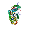
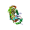
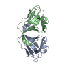
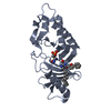
- PDB-4irv: Structure of the Helicobacter pylori CagA Oncogene Bound to the H... -
+ Open data

- Basic information
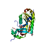
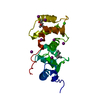
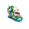
| Entry | Database: PDB / ID: 4irv | ||||||
|---|---|---|---|---|---|---|---|
| Title | Structure of the Helicobacter pylori CagA Oncogene Bound to the Human Tumor Suppressor Apoptosis-stimulating Protein of p53-2 | ||||||
 Components Components |
| ||||||
 Keywords Keywords |  PROTEIN BINDING / Virulence factor and tumor suppressor PROTEIN BINDING / Virulence factor and tumor suppressor | ||||||
| Function / homology |  Function and homology information Function and homology informationtoxin transmembrane transporter activity / TP53 Regulates Transcription of Death Receptors and Ligands / Activation of PUMA and translocation to mitochondria / Regulation of TP53 Activity through Association with Co-factors / positive regulation of execution phase of apoptosis / TP53 regulates transcription of several additional cell death genes whose specific roles in p53-dependent apoptosis remain uncertain / TP53 Regulates Transcription of Genes Involved in Cytochrome C Release / intrinsic apoptotic signaling pathway by p53 class mediator / negative regulation of cell cycle / NF-kappaB binding ...toxin transmembrane transporter activity / TP53 Regulates Transcription of Death Receptors and Ligands / Activation of PUMA and translocation to mitochondria / Regulation of TP53 Activity through Association with Co-factors / positive regulation of execution phase of apoptosis / TP53 regulates transcription of several additional cell death genes whose specific roles in p53-dependent apoptosis remain uncertain / TP53 Regulates Transcription of Genes Involved in Cytochrome C Release / intrinsic apoptotic signaling pathway by p53 class mediator / negative regulation of cell cycle / NF-kappaB binding / SH3 domain binding / p53 binding / cell junction / molecular adaptor activity / cell cycle / perinuclear region of cytoplasm / signal transduction / nucleoplasm / identical protein binding / nucleus / cytosol / cytoplasmSimilarity search - Function | ||||||
| Biological species |   Helicobacter pylori (bacteria) Helicobacter pylori (bacteria) Homo sapiens (human) Homo sapiens (human) | ||||||
| Method | X-RAY DIFFRACTION / SYNCHROTRON / SAD / Resolution: 2.04 Å | ||||||
 Authors Authors | Stebbins, C.E. / Nesic, D. | ||||||
 Citation Citation | Journal: Proc.Natl.Acad.Sci.USA / Year: 2014 Title: Structure of the Helicobacter pylori CagA oncoprotein bound to the human tumor suppressor ASPP2. Authors: Nesic, D. / Buti, L. / Lu, X. / Stebbins, C.E. | ||||||
| History |
|
- Structure visualization
| Structure viewer | Molecule: MolmilJmol/JSmol |
|---|
- Downloads & links
-Download
| PDBx/mmCIF format | 4irv.cif.gz | 191 KB | Display | PDBx/mmCIF format |
|---|---|---|---|---|
| PDB format | pdb4irv.ent.gz | 160.9 KB | Display | PDB format |
| PDBx/mmJSON format | 4irv.json.gz | Tree view | PDBx/mmJSON format | |
| Others |  Other downloads Other downloads |
-Validation report
| Arichive directory | https://data.pdbj.org/pub/pdb/validation_reports/ir/4irvftp://data.pdbj.org/pub/pdb/validation_reports/ir/4irv | HTTPS FTP |
|---|
-Related structure data
| Similar structure data |
|---|


-Links
 PDBj
PDBj


- Assembly
| Deposited unit | 
| |||||||||||||||||||||
|---|---|---|---|---|---|---|---|---|---|---|---|---|---|---|---|---|---|---|---|---|---|---|
| 1 | 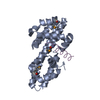
| |||||||||||||||||||||
| 2 | 
| |||||||||||||||||||||
| 3 | 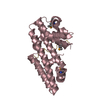
| |||||||||||||||||||||
| 4 | 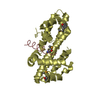
| |||||||||||||||||||||
| Unit cell |
| |||||||||||||||||||||
| Components on special symmetry positions |
|
-Components
| #1: Protein | Mass: 25480.021 Da / Num. of mol.: 4 Source method: isolated from a genetically manipulated source Source: (gene. exp.) Helicobacter pylori (bacteria) / Strain: ATCC 700392 / 26695 / Gene: cagA, cag26, cai, HP_0547 / Production host: Escherichia coli (E. coli) / References: UniProt: P55980#2: Protein | Mass: 6609.313 Da / Num. of mol.: 4 Source method: isolated from a genetically manipulated source Source: (gene. exp.) Homo sapiens (human) / Gene: ASPP2, BBP, TP53BP2 / Production host: Escherichia coli (E. coli) / References: UniProt: Q13625#3: Water | ChemComp-HOH / | Water Mass: 18.015 Da / Num. of mol.: 513 / Source method: isolated from a natural source / Formula: H2O Mass: 18.015 Da / Num. of mol.: 513 / Source method: isolated from a natural source / Formula: H2O |
|---|
-Experimental details
-Experiment
| Experiment | Method: X-RAY DIFFRACTION |
|---|
- Sample preparation
| Crystal | Density Matthews: 2.53 Å3/Da / Density % sol: 51.29 % |
|---|---|
| Crystal grow | Method: vapor diffusion, hanging drop / pH: 8.5 Details: Crystals were grown by vapor diffusion at 25 degrees C using hanging drops formed from mixing a 2ul of the protein complex with 2ul of an equilibration buffer (21% polyethylene glycol (PEG) ...Details: Crystals were grown by vapor diffusion at 25 degrees C using hanging drops formed from mixing a 2ul of the protein complex with 2ul of an equilibration buffer (21% polyethylene glycol (PEG) molecular weight 4 kDa, 200 mM Li2 SO4 , and 100 mM Tris pH 8.5) and 0.6ul of the Silver Bullets additive 43 (Hampton Research HR2-996-43). Significantly higher quality crystals were obtained from selenomethionine-substituted protein complexes and they were used for the final refinement, VAPOR DIFFUSION, HANGING DROP |
-Data collection
| Diffraction source | Source: SYNCHROTRON / Site: NSLS  / Beamline: X29A / Wavelength: 0.97869 Å / Beamline: X29A / Wavelength: 0.97869 Å |
|---|---|
| Detector | Type: ADSC QUANTUM 315 / Detector: CCD |
| Radiation | Protocol: SINGLE WAVELENGTH / Monochromatic (M) / Laue (L): M / Scattering type: x-ray |
| Radiation wavelength | Wavelength: 0.97869 Å / Relative weight: 1 |
| Reflection | Resolution: 2.04→90.75 Å / Num. obs: 80050 |
- Processing
| Software | Name: REFMAC / Version: 5.7.0029 / Classification: refinement | ||||||||||||||||||||||||||||||||||||||||||||||||||||||||||||||||||||||||||||||||||||||||||||||||||||||||||||||||||||||||||||||||||||||||||||||||||||||||||||||||||||||||||
|---|---|---|---|---|---|---|---|---|---|---|---|---|---|---|---|---|---|---|---|---|---|---|---|---|---|---|---|---|---|---|---|---|---|---|---|---|---|---|---|---|---|---|---|---|---|---|---|---|---|---|---|---|---|---|---|---|---|---|---|---|---|---|---|---|---|---|---|---|---|---|---|---|---|---|---|---|---|---|---|---|---|---|---|---|---|---|---|---|---|---|---|---|---|---|---|---|---|---|---|---|---|---|---|---|---|---|---|---|---|---|---|---|---|---|---|---|---|---|---|---|---|---|---|---|---|---|---|---|---|---|---|---|---|---|---|---|---|---|---|---|---|---|---|---|---|---|---|---|---|---|---|---|---|---|---|---|---|---|---|---|---|---|---|---|---|---|---|---|---|---|---|
| Refinement | Method to determine structure: SAD / Resolution: 2.04→90.75 Å / Cor.coef. Fo:Fc: 0.946 / Cor.coef. Fo:Fc free: 0.926 / SU B: 3.95 / SU ML: 0.108 / Cross valid method: THROUGHOUT / ESU R: 0.165 / ESU R Free: 0.157 / Stereochemistry target values: MAXIMUM LIKELIHOOD / Details: HYDROGENS HAVE BEEN ADDED IN THE RIDING POSITIONS
| ||||||||||||||||||||||||||||||||||||||||||||||||||||||||||||||||||||||||||||||||||||||||||||||||||||||||||||||||||||||||||||||||||||||||||||||||||||||||||||||||||||||||||
| Solvent computation | Ion probe radii: 0.8 Å / Shrinkage radii: 0.8 Å / VDW probe radii: 1.2 Å / Solvent model: MASK | ||||||||||||||||||||||||||||||||||||||||||||||||||||||||||||||||||||||||||||||||||||||||||||||||||||||||||||||||||||||||||||||||||||||||||||||||||||||||||||||||||||||||||
| Displacement parameters | Biso mean: 34 Å2
| ||||||||||||||||||||||||||||||||||||||||||||||||||||||||||||||||||||||||||||||||||||||||||||||||||||||||||||||||||||||||||||||||||||||||||||||||||||||||||||||||||||||||||
| Refinement step | Cycle: LAST / Resolution: 2.04→90.75 Å
| ||||||||||||||||||||||||||||||||||||||||||||||||||||||||||||||||||||||||||||||||||||||||||||||||||||||||||||||||||||||||||||||||||||||||||||||||||||||||||||||||||||||||||
| Refine LS restraints |
| ||||||||||||||||||||||||||||||||||||||||||||||||||||||||||||||||||||||||||||||||||||||||||||||||||||||||||||||||||||||||||||||||||||||||||||||||||||||||||||||||||||||||||
| LS refinement shell | Resolution: 2.045→2.098 Å / Total num. of bins used: 20
|
