 Movie
Movie Controller

[English] 日本語
 Yorodumi
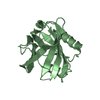
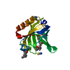
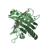
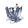
- PDB-4imn: Crystal structure of wild type human Lipocalin PGDS bound with PE... -
+ Open data

- Basic information
| Entry | Database: PDB / ID: 4imn | ||||||
|---|---|---|---|---|---|---|---|
| Title | Crystal structure of wild type human Lipocalin PGDS bound with PEG MME 2000 | ||||||
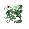
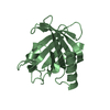
 Components Components | Lipocalin-type prostaglandin-D synthase | ||||||
 Keywords Keywords |  ISOMERASE / Beta barrel / Endoplasmic Reticulum ISOMERASE / Beta barrel / Endoplasmic Reticulum | ||||||
| Function / homology |  Function and homology informationprostaglandin-D synthase / Transcriptional regulation of testis differentiation / prostaglandin-D synthase activity / negative regulation of male germ cell proliferation / regulation of circadian sleep/wake cycle, sleep / Synthesis of Prostaglandins (PG) and Thromboxanes (TX) / cyclooxygenase pathway / retinoid binding / prostaglandin biosynthetic process / mast cell degranulation ...prostaglandin-D synthase / Transcriptional regulation of testis differentiation / prostaglandin-D synthase activity / negative regulation of male germ cell proliferation / regulation of circadian sleep/wake cycle, sleep / Synthesis of Prostaglandins (PG) and Thromboxanes (TX) / cyclooxygenase pathway / retinoid binding / prostaglandin biosynthetic process / mast cell degranulation / rough endoplasmic reticulum / response to glucocorticoid / fatty acid binding / gene expression / nuclear membrane / endoplasmic reticulum membrane / perinuclear region of cytoplasm / Golgi apparatus / extracellular space / extracellular exosome / extracellular region Function and homology informationprostaglandin-D synthase / Transcriptional regulation of testis differentiation / prostaglandin-D synthase activity / negative regulation of male germ cell proliferation / regulation of circadian sleep/wake cycle, sleep / Synthesis of Prostaglandins (PG) and Thromboxanes (TX) / cyclooxygenase pathway / retinoid binding / prostaglandin biosynthetic process / mast cell degranulation ...prostaglandin-D synthase / Transcriptional regulation of testis differentiation / prostaglandin-D synthase activity / negative regulation of male germ cell proliferation / regulation of circadian sleep/wake cycle, sleep / Synthesis of Prostaglandins (PG) and Thromboxanes (TX) / cyclooxygenase pathway / retinoid binding / prostaglandin biosynthetic process / mast cell degranulation / rough endoplasmic reticulum / response to glucocorticoid / fatty acid binding / gene expression / nuclear membrane / endoplasmic reticulum membrane / perinuclear region of cytoplasm / Golgi apparatus / extracellular space / extracellular exosome / extracellular regionSimilarity search - Function | ||||||
| Biological species |  Homo sapiens (human) Homo sapiens (human) | ||||||
| Method | X-RAY DIFFRACTION / SYNCHROTRON / MOLECULAR REPLACEMENT / Resolution: 2.09 Å | ||||||
 Authors Authors | Lim, S.M. / Chen, D. / Teo, H. / Roos, A. / Nyman, T. / Tresaugues, L. / Pervushin, K. / Nordlund, P. | ||||||
 Citation Citation | Journal: J.Lipid Res. / Year: 2013 Title: Structural and dynamic insights into substrate binding and catalysis of human lipocalin prostaglandin D synthase. Authors: Lim, S.M. / Chen, D. / Teo, H. / Roos, A. / Jansson, A.E. / Nyman, T. / Tresaugues, L. / Pervushin, K. / Nordlund, P. | ||||||
| History |
|
- Structure visualization
| Structure viewer | Molecule: MolmilJmol/JSmol |
|---|
- Downloads & links
-Download
| PDBx/mmCIF format | 4imn.cif.gz | 46.6 KB | Display | PDBx/mmCIF format |
|---|---|---|---|---|
| PDB format | pdb4imn.ent.gz | 31.5 KB | Display | PDB format |
| PDBx/mmJSON format | 4imn.json.gz | Tree view | PDBx/mmJSON format | |
| Others |  Other downloads Other downloads |
-Validation report
| Arichive directory | https://data.pdbj.org/pub/pdb/validation_reports/im/4imnftp://data.pdbj.org/pub/pdb/validation_reports/im/4imn | HTTPS FTP |
|---|
-Related structure data
| Related structure data | 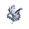 2wwpC  4imoC 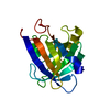 2cztS C: citing same article ( S: Starting model for refinement |
|---|---|
| Similar structure data |


-Links
 PDBj
PDBj


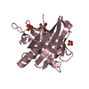
- Assembly
| Deposited unit | 
| ||||||||
|---|---|---|---|---|---|---|---|---|---|
| 1 |
| ||||||||
| Unit cell |
|
-Components
| #1: Protein | Mass: 19748.145 Da / Num. of mol.: 1 Source method: isolated from a genetically manipulated source Source: (gene. exp.) Homo sapiens (human) / Gene: PDS, PTGDS / Plasmid: pNIC-CH4 / Production host:  Escherichia coli (E. coli) / Strain (production host): Rosetta BL21 / References: UniProt: P41222, prostaglandin-D synthase Escherichia coli (E. coli) / Strain (production host): Rosetta BL21 / References: UniProt: P41222, prostaglandin-D synthase |
|---|---|
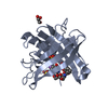
| #2: Chemical | ChemComp-1PG / Polyethylene glycol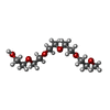  Mass: 252.305 Da / Num. of mol.: 1 / Source method: obtained synthetically / Formula: C11H24O6 Mass: 252.305 Da / Num. of mol.: 1 / Source method: obtained synthetically / Formula: C11H24O6 |
| #3: Water | ChemComp-HOH / Water Mass: 18.015 Da / Num. of mol.: 59 / Source method: isolated from a natural source / Formula: H2O Mass: 18.015 Da / Num. of mol.: 59 / Source method: isolated from a natural source / Formula: H2O |
-Experimental details
-Experiment
| Experiment | Method: X-RAY DIFFRACTION / Number of used crystals: 1 |
|---|
- Sample preparation
| Crystal | Density Matthews: 1.9 Å3/Da / Density % sol: 35.2 % |
|---|---|
| Crystal grow | Temperature: 277 K / Method: vapor diffusion, hanging drop / pH: 6.5 Details: 0.1M potassium thiocyanate, 30% PEG MME 2000, pH 6.5, VAPOR DIFFUSION, HANGING DROP, temperature 277K |
-Data collection
| Diffraction | Mean temperature: 199 K |
|---|---|
| Diffraction source | Source: SYNCHROTRON / Site: NSRRC  / Beamline: BL13B1 / Wavelength: 0.97622 Å / Beamline: BL13B1 / Wavelength: 0.97622 Å |
| Detector | Type: ADSC QUANTUM 315 / Detector: CCD / Date: Sep 30, 2011 |
| Radiation | Monochromator: LN2-cooled, fixed-exit double crystal monochromator Protocol: SINGLE WAVELENGTH / Monochromatic (M) / Laue (L): M / Scattering type: x-ray |
| Radiation wavelength | Wavelength: 0.97622 Å / Relative weight: 1 |
| Reflection | Resolution: 2.09→23.46 Å / Num. all: 8901 / Num. obs: 8428 / % possible obs: 94.6 % / Observed criterion σ(F): 0 / Observed criterion σ(I): -3 / Redundancy: 4.6 % / Biso Wilson estimate: 35.5 Å2 / Rmerge(I) obs: 0.073 / Net I/σ(I): 18.6 |
| Reflection shell | Resolution: 2.09→2.16 Å / Redundancy: 3.4 % / Rmerge(I) obs: 0.345 / Mean I/σ(I) obs: 3.23 / Num. unique all: 824 / % possible all: 94.7 |
- Processing
| Software |
| ||||||||||||||||||||||||||||||||||||||||||||||||||||||||||||
|---|---|---|---|---|---|---|---|---|---|---|---|---|---|---|---|---|---|---|---|---|---|---|---|---|---|---|---|---|---|---|---|---|---|---|---|---|---|---|---|---|---|---|---|---|---|---|---|---|---|---|---|---|---|---|---|---|---|---|---|---|---|
| Refinement | Method to determine structure: MOLECULAR REPLACEMENT Starting model: 2CZT Resolution: 2.09→23.46 Å / Cor.coef. Fo:Fc: 0.962 / Cor.coef. Fo:Fc free: 0.935 / SU B: 4.316 / SU ML: 0.113 / Cross valid method: THROUGHOUT / σ(F): 0 / σ(I): -3 / ESU R: 0.23 / ESU R Free: 0.179 / Stereochemistry target values: MAXIMUM LIKELIHOOD / Details: HYDROGENS HAVE BEEN ADDED IN THE RIDING POSITIONS
| ||||||||||||||||||||||||||||||||||||||||||||||||||||||||||||
| Solvent computation | Ion probe radii: 0.8 Å / Shrinkage radii: 0.8 Å / VDW probe radii: 1.2 Å / Solvent model: MASK | ||||||||||||||||||||||||||||||||||||||||||||||||||||||||||||
| Displacement parameters | Biso mean: 35.449 Å2
| ||||||||||||||||||||||||||||||||||||||||||||||||||||||||||||
| Refinement step | Cycle: LAST / Resolution: 2.09→23.46 Å
| ||||||||||||||||||||||||||||||||||||||||||||||||||||||||||||
| Refine LS restraints |
| ||||||||||||||||||||||||||||||||||||||||||||||||||||||||||||
| LS refinement shell | Resolution: 2.092→2.146 Å / Total num. of bins used: 20
|
