 Movie
Movie Controller

[English] 日本語
 Yorodumi
- PDB-1l6m: Neutrophil Gelatinase-associated Lipocalin is a Novel Bacteriosta... -
+ Open data

- Basic information
| Entry | Database: PDB / ID: 1l6m | ||||||
|---|---|---|---|---|---|---|---|
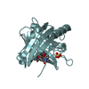
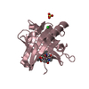
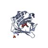
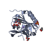
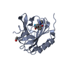
| Title | Neutrophil Gelatinase-associated Lipocalin is a Novel Bacteriostatic Agent that Interferes with Siderophore-mediated Iron Acquisition | ||||||
 Components Components | Neutrophil gelatinase-associated lipocalin | ||||||
 Keywords Keywords |  TRANSPORT PROTEIN / lipocalin / siderophore TRANSPORT PROTEIN / lipocalin / siderophore | ||||||
| Function / homology |  Function and homology information Function and homology informationsiderophore transport / Metal sequestration by antimicrobial proteins / iron ion sequestering activity / enterobactin binding / Iron uptake and transport / specific granule lumen / positive regulation of cold-induced thermogenesis / Interleukin-4 and Interleukin-13 signaling / defense response to bacterium / iron ion binding ...siderophore transport / Metal sequestration by antimicrobial proteins / iron ion sequestering activity / enterobactin binding / Iron uptake and transport / specific granule lumen / positive regulation of cold-induced thermogenesis / Interleukin-4 and Interleukin-13 signaling / defense response to bacterium / iron ion binding / innate immune response / apoptotic process / Neutrophil degranulation / extracellular space / extracellular exosome / extracellular region / identical protein bindingSimilarity search - Function | ||||||
| Biological species |  Homo sapiens (human) Homo sapiens (human) | ||||||
| Method | X-RAY DIFFRACTION / SYNCHROTRON / MOLECULAR REPLACEMENT / Resolution: 2.4 Å | ||||||
 Authors Authors | Goetz, D.H. / Borregaard, N. / Bluhm, M.E. / Raymond, K.N. / Strong, R.K. | ||||||
 Citation Citation | Journal: Mol.Cell / Year: 2002 Title: The Neutrophil Lipocalin NGAL is a Bacteriostatic Agent that Interferes with Siderophore-mediated Iron Acquisition Authors: Goetz, D.H. / Borregaard, N. / Bluhm, M.E. / Raymond, K.N. / Strong, R.K. #1: Journal: Biochemistry / Year: 2000Title: Ligand preference inferred from the structure of Neutrophil Gelatinase Associated Lipocalin (NGAL) Authors: Goetz, D.H. / Willie, S.T. / Armen, R. / Bratt, T. / Borregaard, N. / Strong, R.K. | ||||||
| History |
|
- Structure visualization
| Structure viewer | Molecule: MolmilJmol/JSmol |
|---|
- Downloads & links
-Download
| PDBx/mmCIF format | 1l6m.cif.gz | 121.3 KB | Display | PDBx/mmCIF format |
|---|---|---|---|---|
| PDB format | pdb1l6m.ent.gz | 94.3 KB | Display | PDB format |
| PDBx/mmJSON format | 1l6m.json.gz | Tree view | PDBx/mmJSON format | |
| Others |  Other downloads Other downloads |
-Validation report
| Arichive directory | https://data.pdbj.org/pub/pdb/validation_reports/l6/1l6mftp://data.pdbj.org/pub/pdb/validation_reports/l6/1l6m | HTTPS FTP |
|---|
-Related structure data
| Related structure data |  1qqsS S: Starting model for refinement |
|---|---|
| Similar structure data |


-Links
 PDBj
PDBj

- Assembly
| Deposited unit | 
| ||||||||
|---|---|---|---|---|---|---|---|---|---|
| 1 | 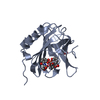
| ||||||||
| 2 | 
| ||||||||
| 3 | 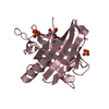
| ||||||||
| Unit cell |
|
-Components
-Protein , 1 types, 3 molecules ABC
| #1: Protein | Mass: 20700.564 Da / Num. of mol.: 3 / Mutation: C87S Source method: isolated from a genetically manipulated source Source: (gene. exp.) Homo sapiens (human) / Production host:  Escherichia coli (E. coli) / References: UniProt: P80188 Escherichia coli (E. coli) / References: UniProt: P80188 |
|---|
-Non-polymers , 5 types, 75 molecules 


| #2: Chemical | Iron Mass: 55.845 Da / Num. of mol.: 3 / Source method: obtained synthetically / Formula: Fe Mass: 55.845 Da / Num. of mol.: 3 / Source method: obtained synthetically / Formula: Fe#3: Chemical | Sulfate Mass: 96.063 Da / Num. of mol.: 3 / Source method: obtained synthetically / Formula: SO4 Mass: 96.063 Da / Num. of mol.: 3 / Source method: obtained synthetically / Formula: SO4#4: Chemical | ChemComp-DBH / 2,3-Dihydroxybenzoic acid Mass: 154.120 Da / Num. of mol.: 5 / Source method: obtained synthetically / Formula: C7H6O4 Mass: 154.120 Da / Num. of mol.: 5 / Source method: obtained synthetically / Formula: C7H6O4#5: Chemical |  Type: L-peptide linking / Mass: 241.197 Da / Num. of mol.: 3 / Source method: obtained synthetically / Formula: C10H11NO6 Type: L-peptide linking / Mass: 241.197 Da / Num. of mol.: 3 / Source method: obtained synthetically / Formula: C10H11NO6#6: Water | ChemComp-HOH / | WaterMass: 18.015 Da / Num. of mol.: 61 / Source method: isolated from a natural source / Formula: H2O |
|---|
-Experimental details
-Experiment
| Experiment | Method: X-RAY DIFFRACTION / Number of used crystals: 1 |
|---|
- Sample preparation
| Crystal | Density Matthews: 3.07 Å3/Da / Density % sol: 59.93 % | |||||||||||||||||||||||||
|---|---|---|---|---|---|---|---|---|---|---|---|---|---|---|---|---|---|---|---|---|---|---|---|---|---|---|
| Crystal grow | Temperature: 298 K / pH: 7 Details: PEG 8000, ammonium sulphate, glycerol, PIPES, pH 7.0, VAPOR DIFFUSION, HANGING DROP, temperature 298.0K, pH 7.00 | |||||||||||||||||||||||||
| Crystal grow | *PLUS pH: 4.5 / Method: vapor diffusion / Details: Goetz, D.H., (2000) Biochemistry, 39, 1935. | |||||||||||||||||||||||||
| Components of the solutions | *PLUS
|
-Data collection
| Diffraction | Mean temperature: 100 K |
|---|---|
| Diffraction source | Source: SYNCHROTRON / Site: ALS  / Beamline: 5.0.1 / Beamline: 5.0.1 |
| Detector | Type: ADSC QUANTUM 4 / Detector: CCD |
| Radiation | Monochromator: SINGLE CRYSTAL, CYLINDRICALLY BENT / Protocol: SINGLE WAVELENGTH / Monochromatic (M) / Laue (L): M / Scattering type: x-ray |
| Radiation wavelength | Relative weight: 1 |
| Reflection | Resolution: 2.4→19.76 Å / Num. obs: 29626 / % possible obs: 96.5 % / Rsym value: 0.057 |
| Reflection | *PLUS Highest resolution: 2.4 Å / Lowest resolution: 20 Å / Num. obs: 30746 / % possible obs: 100 % / Num. measured all: 312664 / Rmerge(I) obs: 0.057 |
| Reflection shell | *PLUS Highest resolution: 2.4 Å / Lowest resolution: 2.44 Å / % possible obs: 100 % / Rmerge(I) obs: 0.51 / Mean I/σ(I) obs: 3.8 |
- Processing
| Software |
| ||||||||||||||||||||||||||||||||||||||||||||||||||||||||||||||||||||||||||||||||||||||||||||||||||||||||||||||||||||||||||||||||||
|---|---|---|---|---|---|---|---|---|---|---|---|---|---|---|---|---|---|---|---|---|---|---|---|---|---|---|---|---|---|---|---|---|---|---|---|---|---|---|---|---|---|---|---|---|---|---|---|---|---|---|---|---|---|---|---|---|---|---|---|---|---|---|---|---|---|---|---|---|---|---|---|---|---|---|---|---|---|---|---|---|---|---|---|---|---|---|---|---|---|---|---|---|---|---|---|---|---|---|---|---|---|---|---|---|---|---|---|---|---|---|---|---|---|---|---|---|---|---|---|---|---|---|---|---|---|---|---|---|---|---|---|
| Refinement | Method to determine structure: MOLECULAR REPLACEMENT Starting model: PDB ENTRY 1QQS Resolution: 2.4→19.76 Å / Cor.coef. Fo:Fc: 0.927 / Cor.coef. Fo:Fc free: 0.914 / SU B: 8.133 / SU ML: 0.192 / Isotropic thermal model: ISOTROPIC AND TLS / Cross valid method: THROUGHOUT / ESU R: 0.362 / ESU R Free: 0.25 / Stereochemistry target values: MAXIMUM LIKELIHOOD / Details: CNS was also used for refinement.
| ||||||||||||||||||||||||||||||||||||||||||||||||||||||||||||||||||||||||||||||||||||||||||||||||||||||||||||||||||||||||||||||||||
| Solvent computation | Ion probe radii: 0.8 Å / Shrinkage radii: 0.8 Å / VDW probe radii: 1.4 Å / Solvent model: BABINET MODEL WITH MASK | ||||||||||||||||||||||||||||||||||||||||||||||||||||||||||||||||||||||||||||||||||||||||||||||||||||||||||||||||||||||||||||||||||
| Displacement parameters | Biso mean: 38.184 Å2
| ||||||||||||||||||||||||||||||||||||||||||||||||||||||||||||||||||||||||||||||||||||||||||||||||||||||||||||||||||||||||||||||||||
| Refinement step | Cycle: LAST / Resolution: 2.4→19.76 Å
| ||||||||||||||||||||||||||||||||||||||||||||||||||||||||||||||||||||||||||||||||||||||||||||||||||||||||||||||||||||||||||||||||||
| Refine LS restraints |
| ||||||||||||||||||||||||||||||||||||||||||||||||||||||||||||||||||||||||||||||||||||||||||||||||||||||||||||||||||||||||||||||||||
| LS refinement shell | Resolution: 2.399→2.461 Å / Total num. of bins used: 20 /
| ||||||||||||||||||||||||||||||||||||||||||||||||||||||||||||||||||||||||||||||||||||||||||||||||||||||||||||||||||||||||||||||||||
| Refinement TLS params. | Method: refined / Refine-ID: X-RAY DIFFRACTION
| ||||||||||||||||||||||||||||||||||||||||||||||||||||||||||||||||||||||||||||||||||||||||||||||||||||||||||||||||||||||||||||||||||
| Refinement TLS group |
| ||||||||||||||||||||||||||||||||||||||||||||||||||||||||||||||||||||||||||||||||||||||||||||||||||||||||||||||||||||||||||||||||||
| Refinement | *PLUS Rfactor Rfree: 0.271 / Rfactor Rwork: 0.225 | ||||||||||||||||||||||||||||||||||||||||||||||||||||||||||||||||||||||||||||||||||||||||||||||||||||||||||||||||||||||||||||||||||
| Solvent computation | *PLUS | ||||||||||||||||||||||||||||||||||||||||||||||||||||||||||||||||||||||||||||||||||||||||||||||||||||||||||||||||||||||||||||||||||
| Displacement parameters | *PLUS | ||||||||||||||||||||||||||||||||||||||||||||||||||||||||||||||||||||||||||||||||||||||||||||||||||||||||||||||||||||||||||||||||||
| Refine LS restraints | *PLUS
| ||||||||||||||||||||||||||||||||||||||||||||||||||||||||||||||||||||||||||||||||||||||||||||||||||||||||||||||||||||||||||||||||||
| LS refinement shell | *PLUS Highest resolution: 2.4 Å / Lowest resolution: 2.46 Å / Rfactor Rfree: 0.359 / Rfactor Rwork: 0.25 |
