 Movie
Movie Controller

[English] 日本語
 Yorodumi
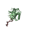
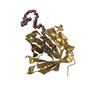
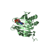
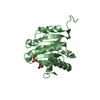
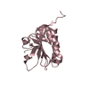
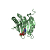
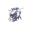
- PDB-4oru: Three-dimensional structure of the C65A mutant of Human lipocalin... -
+ Open data

- Basic information
| Entry | Database: PDB / ID: 4oru | ||||||
|---|---|---|---|---|---|---|---|
| Title | Three-dimensional structure of the C65A mutant of Human lipocalin-type Prostaglandin D Synthase holo-form second space group | ||||||
 Components Components | Prostaglandin-H2 D-isomerase | ||||||
 Keywords Keywords |  ISOMERASE / LIPOCALIN-TYPE PROSTAGLANDIN-D SYNTHASE / PEG ISOMERASE / LIPOCALIN-TYPE PROSTAGLANDIN-D SYNTHASE / PEG | ||||||
| Function / homology |  Function and homology informationprostaglandin-D synthase / Transcriptional regulation of testis differentiation / prostaglandin-D synthase activity / negative regulation of male germ cell proliferation / regulation of circadian sleep/wake cycle, sleep / Synthesis of Prostaglandins (PG) and Thromboxanes (TX) / cyclooxygenase pathway / retinoid binding / prostaglandin biosynthetic process / mast cell degranulation ...prostaglandin-D synthase / Transcriptional regulation of testis differentiation / prostaglandin-D synthase activity / negative regulation of male germ cell proliferation / regulation of circadian sleep/wake cycle, sleep / Synthesis of Prostaglandins (PG) and Thromboxanes (TX) / cyclooxygenase pathway / retinoid binding / prostaglandin biosynthetic process / mast cell degranulation / rough endoplasmic reticulum / response to glucocorticoid / fatty acid binding / gene expression / nuclear membrane / endoplasmic reticulum membrane / perinuclear region of cytoplasm / Golgi apparatus / extracellular space / extracellular exosome / extracellular region Function and homology informationprostaglandin-D synthase / Transcriptional regulation of testis differentiation / prostaglandin-D synthase activity / negative regulation of male germ cell proliferation / regulation of circadian sleep/wake cycle, sleep / Synthesis of Prostaglandins (PG) and Thromboxanes (TX) / cyclooxygenase pathway / retinoid binding / prostaglandin biosynthetic process / mast cell degranulation ...prostaglandin-D synthase / Transcriptional regulation of testis differentiation / prostaglandin-D synthase activity / negative regulation of male germ cell proliferation / regulation of circadian sleep/wake cycle, sleep / Synthesis of Prostaglandins (PG) and Thromboxanes (TX) / cyclooxygenase pathway / retinoid binding / prostaglandin biosynthetic process / mast cell degranulation / rough endoplasmic reticulum / response to glucocorticoid / fatty acid binding / gene expression / nuclear membrane / endoplasmic reticulum membrane / perinuclear region of cytoplasm / Golgi apparatus / extracellular space / extracellular exosome / extracellular regionSimilarity search - Function | ||||||
| Biological species |  Homo sapiens (human) Homo sapiens (human) | ||||||
| Method | X-RAY DIFFRACTION / SYNCHROTRON / MOLECULAR REPLACEMENT / Resolution: 1.55 Å | ||||||
 Authors Authors | Perduca, M. / Bovi, M. / Bertinelli, M. / Bertini, E. / Destefanis, L. / Carrizo, M.E. / Capaldi, S. / Monaco, H.L. | ||||||
 Citation Citation | Journal: Acta Crystallogr.,Sect.D / Year: 2014 Title: High-resolution structures of mutants of residues that affect access to the ligand-binding cavity of human lipocalin-type prostaglandin D synthase. Authors: Perduca, M. / Bovi, M. / Bertinelli, M. / Bertini, E. / Destefanis, L. / Carrizo, M.E. / Capaldi, S. / Monaco, H.L. | ||||||
| History |
|
- Structure visualization
| Structure viewer | Molecule: MolmilJmol/JSmol |
|---|
- Downloads & links
-Download
| PDBx/mmCIF format | 4oru.cif.gz | 142.2 KB | Display | PDBx/mmCIF format |
|---|---|---|---|---|
| PDB format | pdb4oru.ent.gz | 110.1 KB | Display | PDB format |
| PDBx/mmJSON format | 4oru.json.gz | Tree view | PDBx/mmJSON format | |
| Others |  Other downloads Other downloads |
-Validation report
| Arichive directory | https://data.pdbj.org/pub/pdb/validation_reports/or/4oruftp://data.pdbj.org/pub/pdb/validation_reports/or/4oru | HTTPS FTP |
|---|
-Related structure data
| Related structure data | 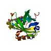 4orrSC 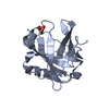 4orsC  4orwC 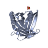 4orxC  4oryC  4os0C  4os3C  4os8C S: Starting model for refinement C: citing same article ( |
|---|---|
| Similar structure data |


-Links
 PDBj
PDBj


- Assembly
| Deposited unit | 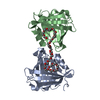
| ||||||||
|---|---|---|---|---|---|---|---|---|---|
| 1 | 
| ||||||||
| 2 | 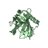
| ||||||||
| 3 |
| ||||||||
| Unit cell |
|
-Components
| #1: Protein | Mass: 21017.717 Da / Num. of mol.: 2 / Mutation: C65A Source method: isolated from a genetically manipulated source Source: (gene. exp.) Homo sapiens (human) / Gene: PTGDS, PDS / Production host:  Escherichia coli (E. coli) / References: UniProt: P41222, prostaglandin-D synthase Escherichia coli (E. coli) / References: UniProt: P41222, prostaglandin-D synthase#2: Chemical | ChemComp-PEU / | Polyethylene glycol  Mass: 1221.461 Da / Num. of mol.: 1 / Source method: obtained synthetically / Formula: C55H112O28 / Comment: precipitant*YM Mass: 1221.461 Da / Num. of mol.: 1 / Source method: obtained synthetically / Formula: C55H112O28 / Comment: precipitant*YM#3: Water | ChemComp-HOH / | Water Mass: 18.015 Da / Num. of mol.: 84 / Source method: isolated from a natural source / Formula: H2O Mass: 18.015 Da / Num. of mol.: 84 / Source method: isolated from a natural source / Formula: H2O |
|---|
-Experimental details
-Experiment
| Experiment | Method: X-RAY DIFFRACTION |
|---|
- Sample preparation
| Crystal | Density Matthews: 1.82 Å3/Da / Density % sol: 32.24 % |
|---|---|
| Crystal grow | Temperature: 293 K / Method: vapor diffusion, hanging drop / pH: 7.5 Details: 30% PEG 4000, 0.2M Ammonium Sulphate, pH 7.5, VAPOR DIFFUSION, HANGING DROP, temperature 293K |
-Data collection
| Diffraction | Mean temperature: 100 K |
|---|---|
| Diffraction source | Source: SYNCHROTRON / Site: ESRF  / Beamline: ID29 / Wavelength: 0.98 Å / Beamline: ID29 / Wavelength: 0.98 Å |
| Detector | Type: DECTRIS PILATUS 6M-F / Detector: PIXEL / Date: Jul 5, 2013 |
| Radiation | Monochromator: Si(111) / Protocol: SINGLE WAVELENGTH / Monochromatic (M) / Laue (L): M / Scattering type: x-ray |
| Radiation wavelength | Wavelength: 0.98 Å / Relative weight: 1 |
| Reflection | Resolution: 1.55→45.56 Å / Num. all: 44356 / Num. obs: 44187 / % possible obs: 99.6 % / Observed criterion σ(F): 1 / Observed criterion σ(I): 1 / Redundancy: 4 % / Rmerge(I) obs: 0.043 / Net I/σ(I): 13.8 |
| Reflection shell | Resolution: 1.55→1.63 Å / Rmerge(I) obs: 0.354 / Mean I/σ(I) obs: 3.3 / % possible all: 99.7 |
- Processing
| Software |
| |||||||||||||||||||||||||||||||||||||||||||||||||||||||||||||||||||||||||||||||||||||||||||||||||||||||||||||||||||||||
|---|---|---|---|---|---|---|---|---|---|---|---|---|---|---|---|---|---|---|---|---|---|---|---|---|---|---|---|---|---|---|---|---|---|---|---|---|---|---|---|---|---|---|---|---|---|---|---|---|---|---|---|---|---|---|---|---|---|---|---|---|---|---|---|---|---|---|---|---|---|---|---|---|---|---|---|---|---|---|---|---|---|---|---|---|---|---|---|---|---|---|---|---|---|---|---|---|---|---|---|---|---|---|---|---|---|---|---|---|---|---|---|---|---|---|---|---|---|---|---|---|
| Refinement | Method to determine structure: MOLECULAR REPLACEMENT Starting model: PDB ENTRY 4ORR Resolution: 1.55→45.565 Å / SU ML: 0.16 / σ(F): 1.34 / Phase error: 23.57 / Stereochemistry target values: ML
| |||||||||||||||||||||||||||||||||||||||||||||||||||||||||||||||||||||||||||||||||||||||||||||||||||||||||||||||||||||||
| Solvent computation | Shrinkage radii: 0.9 Å / VDW probe radii: 1.11 Å / Solvent model: FLAT BULK SOLVENT MODEL | |||||||||||||||||||||||||||||||||||||||||||||||||||||||||||||||||||||||||||||||||||||||||||||||||||||||||||||||||||||||
| Refinement step | Cycle: LAST / Resolution: 1.55→45.565 Å
| |||||||||||||||||||||||||||||||||||||||||||||||||||||||||||||||||||||||||||||||||||||||||||||||||||||||||||||||||||||||
| Refine LS restraints |
| |||||||||||||||||||||||||||||||||||||||||||||||||||||||||||||||||||||||||||||||||||||||||||||||||||||||||||||||||||||||
| LS refinement shell |
| |||||||||||||||||||||||||||||||||||||||||||||||||||||||||||||||||||||||||||||||||||||||||||||||||||||||||||||||||||||||
| Refinement TLS params. | Method: refined / Refine-ID: X-RAY DIFFRACTION
| |||||||||||||||||||||||||||||||||||||||||||||||||||||||||||||||||||||||||||||||||||||||||||||||||||||||||||||||||||||||
| Refinement TLS group |
|
