- PDB-4ijh: Fragment-based Discovery of Protein-Protein Interaction Inhibitor... -
+
Open data
ID or keywords:
Loading...
-
Basic information
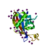
Entry
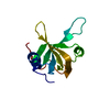
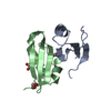
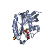
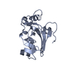
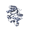
Database: PDB / ID: 4ijh
Title
Fragment-based Discovery of Protein-Protein Interaction Inhibitors of Replication Protein A
Components
Replication protein A 70 kDa DNA-binding subunitDNA replication
Keywords
DNA BINDING PROTEIN/INHIBITOR / OB-Fold / Protein-Protein Interaction / DNA BINDING PROTEIN-INHIBITOR complex
Function / homology
Function and homology information
protein localization to chromosome / DNA replication factor A complex / chromatin-protein adaptor activity / protein localization to site of double-strand break / Removal of the Flap Intermediate / single-stranded telomeric DNA binding / Mismatch repair (MMR) directed by MSH2:MSH3 (MutSbeta) / Mismatch repair (MMR) directed by MSH2:MSH6 (MutSalpha) / Removal of the Flap Intermediate from the C-strand / G-rich strand telomeric DNA binding ...protein localization to chromosome / DNA replication factor A complex / chromatin-protein adaptor activity / protein localization to site of double-strand break / Removal of the Flap Intermediate / single-stranded telomeric DNA binding / Mismatch repair (MMR) directed by MSH2:MSH3 (MutSbeta) / Mismatch repair (MMR) directed by MSH2:MSH6 (MutSalpha) / Removal of the Flap Intermediate from the C-strand / G-rich strand telomeric DNA binding / HDR through Single Strand Annealing (SSA) / Impaired BRCA2 binding to RAD51 / site of DNA damage / Presynaptic phase of homologous DNA pairing and strand exchange / telomere maintenance via telomerase / PCNA-Dependent Long Patch Base Excision Repair / Activation of the pre-replicative complex / HSF1 activation / Regulation of HSF1-mediated heat shock response / mismatch repair / Activation of ATR in response to replication stress / SUMOylation of DNA damage response and repair proteins / telomere maintenance / Translesion synthesis by REV1 / Translesion synthesis by POLK / Gap-filling DNA repair synthesis and ligation in GG-NER / Translesion synthesis by POLI / meiotic cell cycle / nucleotide-excision repair / Recognition of DNA damage by PCNA-containing replication complex / Fanconi Anemia Pathway / Termination of translesion DNA synthesis / double-strand break repair via homologous recombination / Translesion Synthesis by POLH / base-excision repair / HDR through Homologous Recombination (HRR) / G2/M DNA damage checkpoint / Dual Incision in GG-NER / PML body / DNA-templated DNA replication / Meiotic recombination / Formation of Incision Complex in GG-NER / Dual incision in TC-NER / Gap-filling DNA repair synthesis and ligation in TC-NER / single-stranded DNA binding / site of double-strand break / Processing of DNA double-strand break ends / DNA recombination / Regulation of TP53 Activity through Phosphorylation / DNA replication / chromosome, telomeric region / damaged DNA binding / DNA repair / DNA damage response / nucleoplasm / metal ion binding / nucleus Similarity search - Function
Replication factor-A protein 1, N-terminal domain / Replication factor A protein 1 / Replication factor-A protein 1, N-terminal / Replication protein A, OB domain / Replication protein A OB domain / : / Replication factor A, C-terminal / Replication factor-A C terminal domain / OB-fold nucleic acid binding domain, AA-tRNA synthetase-type / OB-fold nucleic acid binding domain ...Replication factor-A protein 1, N-terminal domain / Replication factor A protein 1 / Replication factor-A protein 1, N-terminal / Replication protein A, OB domain / Replication protein A OB domain / : / Replication factor A, C-terminal / Replication factor-A C terminal domain / OB-fold nucleic acid binding domain, AA-tRNA synthetase-type / OB-fold nucleic acid binding domain / Nucleic acid-binding proteins / OB fold (Dihydrolipoamide Acetyltransferase, E2P) / Nucleic acid-binding, OB-fold / Beta Barrel / Mainly Beta Similarity search - Domain/homology
ReplicationproteinA70kDaDNA-bindingsubunit / DNA replication / RP-A p70 / Replication factor A protein 1 / RF-A protein 1 / Single-stranded DNA-binding protein
Mass: 13497.728 Da / Num. of mol.: 1 / Fragment: N-terminal domain (UNP residues 1-120) / Mutation: E7R Source method: isolated from a genetically manipulated source Source: (gene. exp.) Homo sapiens (human) / Gene: REPA1, RPA1, RPA70 / Plasmid: pET15b / Production host: Escherichia coli (E. coli) / Strain (production host): BL21(DE3) / References: UniProt: P27694
In the structure databanks used in Yorodumi, some data are registered as the other names, "COVID-19 virus" and "2019-nCoV". Here are the details of the virus and the list of structure data.
Jan 31, 2019. EMDB accession codes are about to change! (news from PDBe EMDB page)
EMDB accession codes are about to change! (news from PDBe EMDB page)
The allocation of 4 digits for EMDB accession codes will soon come to an end. Whilst these codes will remain in use, new EMDB accession codes will include an additional digit and will expand incrementally as the available range of codes is exhausted. The current 4-digit format prefixed with “EMD-” (i.e. EMD-XXXX) will advance to a 5-digit format (i.e. EMD-XXXXX), and so on. It is currently estimated that the 4-digit codes will be depleted around Spring 2019, at which point the 5-digit format will come into force.
The EM Navigator/Yorodumi systems omit the EMD- prefix.
Related info.:Q: What is EMD? / ID/Accession-code notation in Yorodumi/EM Navigator
Yorodumi is a browser for structure data from EMDB, PDB, SASBDB, etc.
This page is also the successor to EM Navigator detail page, and also detail information page/front-end page for Omokage search.
The word "yorodu" (or yorozu) is an old Japanese word meaning "ten thousand". "mi" (miru) is to see.
Related info.:EMDB / PDB / SASBDB / Comparison of 3 databanks / Yorodumi Search / Aug 31, 2016. New EM Navigator & Yorodumi / Yorodumi Papers / Jmol/JSmol / Function and homology information / Changes in new EM Navigator and Yorodumi
 Movie
Movie Controller

 Yorodumi
 Open data
 Basic information
 Components
 DNA replication
  Keywords
 Function and homology information
 Authors
 Citation
 Structure visualization
 Downloads & links
 Other downloads
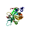


 PDBj
PDBj


 Assembly

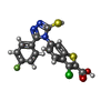
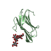
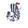
 Mass: 405.854 Da / Num. of mol.: 5 / Source method: obtained synthetically / Formula: C17H9ClFN3O2S2
 Mass: 18.015 Da / Num. of mol.: 133 / Source method: isolated from a natural source / Formula: H2O
 Sample preparation
 / Beamline: 21-ID-D / Wavelength: 0.97857 Å
 Processing