 Movie
Movie Controller

[English] 日本語
 Yorodumi
- PDB-4hly: The complex crystal structure of the DNA binding domain of vIRF-1... -
+ Open data

- Basic information
| Entry | Database: PDB / ID: 4hly | ||||||
|---|---|---|---|---|---|---|---|
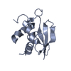
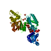
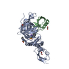
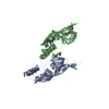
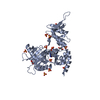
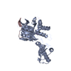
| Title | The complex crystal structure of the DNA binding domain of vIRF-1 from the oncogenic KSHV with DNA | ||||||
 Components Components |
| ||||||
 Keywords Keywords | DNA BINDING PROTEIN/DNA /  helix-turn-helix / DNA BINDING PROTEIN-DNA complex helix-turn-helix / DNA BINDING PROTEIN-DNA complex | ||||||
| Function / homology |  Function and homology information Function and homology information: / protein sequestering activity / symbiont-mediated suppression of host cytoplasmic pattern recognition receptor signaling pathway via inhibition of IRF3 activity / host cell cytoplasm / transcription cis-regulatory region binding / DNA-binding transcription factor activity / protein domain specific binding / host cell nucleus Similarity search - Function | ||||||
| Biological species |   Human herpesvirus 8 Human herpesvirus 8synthetic construct (others) | ||||||
| Method | X-RAY DIFFRACTION / SYNCHROTRON / Resolution: 1.48 Å | ||||||
 Authors Authors | Hew, K. / Venkatachalam, R. | ||||||
 Citation Citation | Journal: Nucleic Acids Res. / Year: 2013 Title: The crystal structure of the DNA-binding domain of vIRF-1 from the oncogenic KSHV reveals a conserved fold for DNA binding and reinforces its role as a transcription factor. Authors: Hew, K. / Dahlroth, S.L. / Venkatachalam, R. / Nasertorabi, F. / Lim, B.T. / Cornvik, T. / Nordlund, P. | ||||||
| History |
|
- Structure visualization
| Structure viewer | Molecule: MolmilJmol/JSmol |
|---|
- Downloads & links
-Download
| PDBx/mmCIF format | 4hly.cif.gz | 77 KB | Display | PDBx/mmCIF format |
|---|---|---|---|---|
| PDB format | pdb4hly.ent.gz | 54.6 KB | Display | PDB format |
| PDBx/mmJSON format | 4hly.json.gz | Tree view | PDBx/mmJSON format | |
| Others |  Other downloads Other downloads |
-Validation report
| Arichive directory | https://data.pdbj.org/pub/pdb/validation_reports/hl/4hlyftp://data.pdbj.org/pub/pdb/validation_reports/hl/4hly | HTTPS FTP |
|---|
-Related structure data


-Links
 PDBj
PDBj


- Assembly
| Deposited unit | 
| ||||||||
|---|---|---|---|---|---|---|---|---|---|
| 1 | 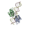
| ||||||||
| Unit cell |
|
-Components
| #1: Protein | Mass: 15309.481 Da / Num. of mol.: 2 / Fragment: DNA binding domain, UNP RESIDUES 88-196 Source method: isolated from a genetically manipulated source Source: (gene. exp.) Human herpesvirus 8 / Gene: ORF K9, vIRF, vIRF-1 / Plasmid: pNIC28-Bsa4 / Production host:  Escherichia coli (E. coli) / Strain (production host): BL21-DE3-Rosetta / References: UniProt: Q77Q82, UniProt: F5HF68*PLUS Escherichia coli (E. coli) / Strain (production host): BL21-DE3-Rosetta / References: UniProt: Q77Q82, UniProt: F5HF68*PLUS#2: DNA chain | Mass: 3688.405 Da / Num. of mol.: 2 / Source method: obtained synthetically / Source: (synth.) synthetic construct (others) #3: Water | ChemComp-HOH / | Water Mass: 18.015 Da / Num. of mol.: 232 / Source method: isolated from a natural source / Formula: H2O Mass: 18.015 Da / Num. of mol.: 232 / Source method: isolated from a natural source / Formula: H2O |
|---|
-Experimental details
-Experiment
| Experiment | Method: X-RAY DIFFRACTION / Number of used crystals: 1 |
|---|
- Sample preparation
| Crystal | Density Matthews: 1.69 Å3/Da / Density % sol: 27.35 % |
|---|---|
| Crystal grow | Temperature: 298 K / Method: vapor diffusion, hanging drop / pH: 4.2 Details: 0.1M phosphate citrate pH 4.2, 40% PEG 300, VAPOR DIFFUSION, HANGING DROP, temperature 298K |
-Data collection
| Diffraction | Mean temperature: 110 K | |||||||||||||||
|---|---|---|---|---|---|---|---|---|---|---|---|---|---|---|---|---|
| Diffraction source | Source: SYNCHROTRON / Site: NSRRC  / Beamline: BL13C1 / Wavelength: 0.96722 Å / Beamline: BL13C1 / Wavelength: 0.96722 Å | |||||||||||||||
| Detector | Type: RAYONIX MX300HE / Detector: CCD / Date: Sep 29, 2010 | |||||||||||||||
| Radiation | Monochromator: Rh Coated Mirror / Protocol: SINGLE WAVELENGTH / Monochromatic (M) / Laue (L): M / Scattering type: x-ray | |||||||||||||||
| Radiation wavelength | Wavelength: 0.96722 Å / Relative weight: 1 | |||||||||||||||
| Reflection twin |
| |||||||||||||||
| Reflection | Resolution: 1.48→50 Å / Num. all: 41661 / Num. obs: 41661 / % possible obs: 99.5 % / Redundancy: 3.8 % / Rmerge(I) obs: 0.106 | |||||||||||||||
| Reflection shell | Highest resolution: 1.48 Å / Redundancy: 3.8 % / Mean I/σ(I) obs: 2.3 / Num. unique all: 41661 / Rsym value: 0.507 / % possible all: 92.8 |
- Processing
| Software |
| ||||||||||||||||||||||||||||||||||||||||||||||||||||||||||||||||||||||||||||||||||||||||||||||||||||||||||||||||||||||||||||||||||||||||||||||||||||||||||||||||||||||||||
|---|---|---|---|---|---|---|---|---|---|---|---|---|---|---|---|---|---|---|---|---|---|---|---|---|---|---|---|---|---|---|---|---|---|---|---|---|---|---|---|---|---|---|---|---|---|---|---|---|---|---|---|---|---|---|---|---|---|---|---|---|---|---|---|---|---|---|---|---|---|---|---|---|---|---|---|---|---|---|---|---|---|---|---|---|---|---|---|---|---|---|---|---|---|---|---|---|---|---|---|---|---|---|---|---|---|---|---|---|---|---|---|---|---|---|---|---|---|---|---|---|---|---|---|---|---|---|---|---|---|---|---|---|---|---|---|---|---|---|---|---|---|---|---|---|---|---|---|---|---|---|---|---|---|---|---|---|---|---|---|---|---|---|---|---|---|---|---|---|---|---|---|
| Refinement | Resolution: 1.48→19.53 Å / Cor.coef. Fo:Fc: 0.959 / Cor.coef. Fo:Fc free: 0.942 / SU B: 1.216 / SU ML: 0.05 / Cross valid method: THROUGHOUT / ESU R: 0.019 / ESU R Free: 0.019 / Stereochemistry target values: MAXIMUM LIKELIHOOD / Details: HYDROGENS HAVE BEEN USED IF PRESENT IN THE INPUT
| ||||||||||||||||||||||||||||||||||||||||||||||||||||||||||||||||||||||||||||||||||||||||||||||||||||||||||||||||||||||||||||||||||||||||||||||||||||||||||||||||||||||||||
| Solvent computation | Ion probe radii: 0.8 Å / Shrinkage radii: 0.8 Å / VDW probe radii: 1.2 Å / Solvent model: MASK | ||||||||||||||||||||||||||||||||||||||||||||||||||||||||||||||||||||||||||||||||||||||||||||||||||||||||||||||||||||||||||||||||||||||||||||||||||||||||||||||||||||||||||
| Displacement parameters | Biso mean: 21.589 Å2
| ||||||||||||||||||||||||||||||||||||||||||||||||||||||||||||||||||||||||||||||||||||||||||||||||||||||||||||||||||||||||||||||||||||||||||||||||||||||||||||||||||||||||||
| Refinement step | Cycle: LAST / Resolution: 1.48→19.53 Å
| ||||||||||||||||||||||||||||||||||||||||||||||||||||||||||||||||||||||||||||||||||||||||||||||||||||||||||||||||||||||||||||||||||||||||||||||||||||||||||||||||||||||||||
| Refine LS restraints |
| ||||||||||||||||||||||||||||||||||||||||||||||||||||||||||||||||||||||||||||||||||||||||||||||||||||||||||||||||||||||||||||||||||||||||||||||||||||||||||||||||||||||||||
| LS refinement shell | Resolution: 1.476→1.514 Å / Total num. of bins used: 20
|
