 Movie
Movie Controller

[English] 日本語
 Yorodumi
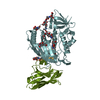
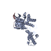
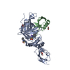
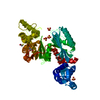
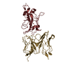
- PDB-6jsz: BACE2 xaperone complex with N-{3-[(5R)-3-amino-5-methyl-9,9-dioxo... -
+ Open data

- Basic information
| Entry | Database: PDB / ID: 6jsz | ||||||
|---|---|---|---|---|---|---|---|
| Title | BACE2 xaperone complex with N-{3-[(5R)-3-amino-5-methyl-9,9-dioxo-2,9lambda6-dithia-4-azaspiro[5.5]undec-3-en-5-yl]-4-fluorophenyl}-5-(fluoromethoxy)pyrazine-2-carboxamide | ||||||
 Components Components |
| ||||||
 Keywords Keywords | HYDROLASE/IMMUNE SYSTEM /  BACE2 / HYDROLASE / HYDROLASE-IMMUNE SYSTEM complex BACE2 / HYDROLASE / HYDROLASE-IMMUNE SYSTEM complex | ||||||
| Function / homology |  Function and homology informationmemapsin 1 / negative regulation of amyloid precursor protein biosynthetic process / melanosome membrane / melanosome organization / peptide hormone processing / membrane protein ectodomain proteolysis / amyloid-beta metabolic process / trans-Golgi network / protein processing / glucose homeostasis ...memapsin 1 / negative regulation of amyloid precursor protein biosynthetic process / melanosome membrane / melanosome organization / peptide hormone processing / membrane protein ectodomain proteolysis / amyloid-beta metabolic process / trans-Golgi network / protein processing / glucose homeostasis / aspartic-type endopeptidase activity / endosome / Golgi apparatus / endoplasmic reticulum / proteolysis / membrane / plasma membrane Function and homology informationmemapsin 1 / negative regulation of amyloid precursor protein biosynthetic process / melanosome membrane / melanosome organization / peptide hormone processing / membrane protein ectodomain proteolysis / amyloid-beta metabolic process / trans-Golgi network / protein processing / glucose homeostasis ...memapsin 1 / negative regulation of amyloid precursor protein biosynthetic process / melanosome membrane / melanosome organization / peptide hormone processing / membrane protein ectodomain proteolysis / amyloid-beta metabolic process / trans-Golgi network / protein processing / glucose homeostasis / aspartic-type endopeptidase activity / endosome / Golgi apparatus / endoplasmic reticulum / proteolysis / membrane / plasma membraneSimilarity search - Function | ||||||
| Biological species |  Homo sapiens (human) Homo sapiens (human) | ||||||
| Method | X-RAY DIFFRACTION / SYNCHROTRON / MOLECULAR REPLACEMENT / Resolution: 1.53 Å | ||||||
 Authors Authors | Fujimoto, K. / Matsuoka, E. / Asada, N. / Tadano, G. / Yamamoto, T. / Nakahara, K. / Fuchino, K. / Ito, H. / Kanegawa, N. / Moechars, D. ...Fujimoto, K. / Matsuoka, E. / Asada, N. / Tadano, G. / Yamamoto, T. / Nakahara, K. / Fuchino, K. / Ito, H. / Kanegawa, N. / Moechars, D. / Gijsen, H.J.M. / Kusakabe, K.I. | ||||||
 Citation Citation | Journal: J.Med.Chem. / Year: 2019 Title: Structure-Based Design of Selective beta-Site Amyloid Precursor Protein Cleaving Enzyme 1 (BACE1) Inhibitors: Targeting the Flap to Gain Selectivity over BACE2. Authors: Fujimoto, K. / Matsuoka, E. / Asada, N. / Tadano, G. / Yamamoto, T. / Nakahara, K. / Fuchino, K. / Ito, H. / Kanegawa, N. / Moechars, D. / Gijsen, H.J.M. / Kusakabe, K.I. | ||||||
| History |
|
- Structure visualization
| Structure viewer | Molecule: MolmilJmol/JSmol |
|---|
- Downloads & links
-Download
| PDBx/mmCIF format | 6jsz.cif.gz | 115.2 KB | Display | PDBx/mmCIF format |
|---|---|---|---|---|
| PDB format | pdb6jsz.ent.gz | 84.2 KB | Display | PDB format |
| PDBx/mmJSON format | 6jsz.json.gz | Tree view | PDBx/mmJSON format | |
| Others |  Other downloads Other downloads |
-Validation report
| Arichive directory | https://data.pdbj.org/pub/pdb/validation_reports/js/6jszftp://data.pdbj.org/pub/pdb/validation_reports/js/6jsz | HTTPS FTP |
|---|
-Related structure data
| Related structure data | 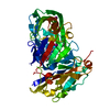 6jseC 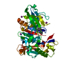 6jsfC  6jsgC  6jsnC 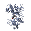 3zkgS C: citing same article ( S: Starting model for refinement |
|---|---|
| Similar structure data |


-Links
 PDBj
PDBj

- Assembly
| Deposited unit | 
| ||||||||
|---|---|---|---|---|---|---|---|---|---|
| 1 |
| ||||||||
| Unit cell |
|
-Components
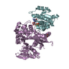
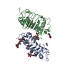
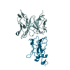
| #1: Protein | / Aspartic-like protease 56 kDa / Aspartyl protease 1 / Asp 1 / Beta-site amyloid precursor protein ...Aspartic-like protease 56 kDa / Aspartyl protease 1 / Asp 1 / Beta-site amyloid precursor protein cleaving enzyme 2 / Beta-site APP cleaving enzyme 2 / Down region aspartic protease / DRAP / Memapsin-1 / Membrane-associated aspartic protease 1 / Theta-secretase Mass: 42105.391 Da / Num. of mol.: 1 / Mutation: AEPLC, ALP56, ASP21 Source method: isolated from a genetically manipulated source Source: (gene. exp.) Homo sapiens (human) / Gene: BACE2, AEPLC, ALP56, ASP21, CDA13, UNQ418/PRO852 / Production host:  Escherichia coli (E. coli) / References: UniProt: Q9Y5Z0, memapsin 1 Escherichia coli (E. coli) / References: UniProt: Q9Y5Z0, memapsin 1 |
|---|---|
| #2: Protein | Mass: 11769.089 Da / Num. of mol.: 1 / Source method: obtained synthetically / Source: (synth.) Homo sapiens (human) |
| #3: Chemical | ChemComp-C7O / 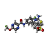  Mass: 511.565 Da / Num. of mol.: 1 / Source method: obtained synthetically / Formula: C21H23F2N5O4S2 Mass: 511.565 Da / Num. of mol.: 1 / Source method: obtained synthetically / Formula: C21H23F2N5O4S2 |
| #4: Chemical | ChemComp-CL / Chloride  Mass: 35.453 Da / Num. of mol.: 1 / Source method: obtained synthetically / Formula: Cl Mass: 35.453 Da / Num. of mol.: 1 / Source method: obtained synthetically / Formula: Cl |
| #5: Water | ChemComp-HOH / Water Mass: 18.015 Da / Num. of mol.: 299 / Source method: isolated from a natural source / Formula: H2O Mass: 18.015 Da / Num. of mol.: 299 / Source method: isolated from a natural source / Formula: H2O |
-Experimental details
-Experiment
| Experiment | Method: X-RAY DIFFRACTION / Number of used crystals: 1 |
|---|
- Sample preparation
| Crystal | Density Matthews: 2.31 Å3/Da / Density % sol: 46.76 % / Description: NONE |
|---|---|
| Crystal grow | Temperature: 293.15 K / Method: vapor diffusion, sitting drop / pH: 8 / Details: 0.1M TRIS pH 8.0, 25% PEG 3350, pH 8.0 |
-Data collection
| Diffraction | Mean temperature: 100 K / Serial crystal experiment: N |
|---|---|
| Diffraction source | Source: SYNCHROTRON / Site: SLS  / Beamline: X06DA / Wavelength: 1 Å / Beamline: X06DA / Wavelength: 1 Å |
| Detector | Type: DECTRIS EIGER X 16M / Detector: PIXEL / Date: Jun 1, 2016 |
| Radiation | Protocol: SINGLE WAVELENGTH / Monochromatic (M) / Laue (L): M / Scattering type: x-ray |
| Radiation wavelength | Wavelength: 1 Å / Relative weight: 1 |
| Reflection | Resolution: 1.53→61.51 Å / Num. obs: 78375 / % possible obs: 99.5 % / Redundancy: 4.1 % / Rrim(I) all: 0.04 / Rsym value: 0.035 / Net I/σ(I): 19.09 |
| Reflection shell | Resolution: 1.53→1.78 Å / Redundancy: 4.1 % / Num. unique obs: 28173 / Rrim(I) all: 0.563 / Rsym value: 0.49 / % possible all: 99.1 |
- Processing
| Software |
| ||||||||||||||||||||||||||||||||||||||||||||||||||||||||||||
|---|---|---|---|---|---|---|---|---|---|---|---|---|---|---|---|---|---|---|---|---|---|---|---|---|---|---|---|---|---|---|---|---|---|---|---|---|---|---|---|---|---|---|---|---|---|---|---|---|---|---|---|---|---|---|---|---|---|---|---|---|---|
| Refinement | Method to determine structure: MOLECULAR REPLACEMENT Starting model: 3ZKG Resolution: 1.53→61.51 Å / Cor.coef. Fo:Fc: 0.955 / Cor.coef. Fo:Fc free: 0.95 / SU B: 2.251 / SU ML: 0.077 / Cross valid method: THROUGHOUT / σ(F): 0 / ESU R: 0.091 / ESU R Free: 0.085 Details: HYDROGENS HAVE BEEN ADDED IN THE RIDING POSITIONS U VALUES : REFINED INDIVIDUALLY
| ||||||||||||||||||||||||||||||||||||||||||||||||||||||||||||
| Solvent computation | Ion probe radii: 0.8 Å / Shrinkage radii: 0.8 Å / VDW probe radii: 1.2 Å | ||||||||||||||||||||||||||||||||||||||||||||||||||||||||||||
| Displacement parameters | Biso max: 84.57 Å2 / Biso mean: 28.96 Å2 / Biso min: 15.92 Å2
| ||||||||||||||||||||||||||||||||||||||||||||||||||||||||||||
| Refinement step | Cycle: final / Resolution: 1.53→61.51 Å
| ||||||||||||||||||||||||||||||||||||||||||||||||||||||||||||
| Refine LS restraints |
| ||||||||||||||||||||||||||||||||||||||||||||||||||||||||||||
| LS refinement shell | Resolution: 1.531→1.571 Å / Rfactor Rfree error: 0 / Total num. of bins used: 20
|
