 Movie
Movie Controller


+ Open data

- Basic information
| Entry | Database: PDB / ID: 4han | ||||||
|---|---|---|---|---|---|---|---|
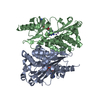
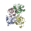
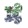
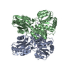
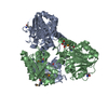
| Title | Crystal structure of Galectin 8 with NDP52 peptide | ||||||
 Components Components |
| ||||||
 Keywords Keywords | SUGAR BINDING PROTEIN /  Autophagy / innate immunity / carbohydrate recognition domain (CRD) / autophagy adapter molecule / NAD binding / NDP52 peoptide binding / Cytosol Autophagy / innate immunity / carbohydrate recognition domain (CRD) / autophagy adapter molecule / NAD binding / NDP52 peoptide binding / Cytosol | ||||||
| Function / homology |  Function and homology information Function and homology informationlymphatic endothelial cell migration / xenophagy / positive regulation of autophagosome maturation / response to type II interferon / autophagosome membrane / autophagosome / viral process / PML body / cellular response to virus / integrin binding ...lymphatic endothelial cell migration / xenophagy / positive regulation of autophagosome maturation / response to type II interferon / autophagosome membrane / autophagosome / viral process / PML body / cellular response to virus / integrin binding / cytoplasmic vesicle / carbohydrate binding / cytoskeleton / intracellular membrane-bounded organelle / perinuclear region of cytoplasm / protein homodimerization activity / extracellular space / membrane / metal ion binding / nucleus / cytosol / cytoplasmSimilarity search - Function | ||||||
| Biological species |  Homo sapiens (human) Homo sapiens (human) | ||||||
| Method | X-RAY DIFFRACTION / SYNCHROTRON / MOLECULAR REPLACEMENT / Resolution: 2.551 Å | ||||||
 Authors Authors | Kim, B.-W. / Hong, S.B. / Kim, J.H. / Kwon, D.H. / Song, H.K. | ||||||
 Citation Citation | Journal: Nat Commun / Year: 2013 Title: Structural basis for recognition of autophagic receptor NDP52 by the sugar receptor galectin-8. Authors: Kim, B.W. / Hong, S.B. / Kim, J.H. / Kwon, D.H. / Song, H.K. | ||||||
| History |
|
- Structure visualization
| Structure viewer | Molecule: MolmilJmol/JSmol |
|---|
- Downloads & links
-Download
| PDBx/mmCIF format | 4han.cif.gz | 259.8 KB | Display | PDBx/mmCIF format |
|---|---|---|---|---|
| PDB format | pdb4han.ent.gz | 211.8 KB | Display | PDB format |
| PDBx/mmJSON format | 4han.json.gz | Tree view | PDBx/mmJSON format | |
| Others |  Other downloads Other downloads |
-Validation report
| Arichive directory | https://data.pdbj.org/pub/pdb/validation_reports/ha/4hanftp://data.pdbj.org/pub/pdb/validation_reports/ha/4han | HTTPS FTP |
|---|
-Related structure data
| Similar structure data |
|---|


-Links
 PDBj
PDBj
- Assembly
| Deposited unit | 
| ||||||||
|---|---|---|---|---|---|---|---|---|---|
| 1 |
| ||||||||
| Unit cell |
|
-Components
| #1: Protein | Mass: 33222.207 Da / Num. of mol.: 2 / Fragment: UNP residues 1-155. 184-317 Source method: isolated from a genetically manipulated source Source: (gene. exp.) Homo sapiens (human) / Production host:  Escherichia coli (E. coli) / References: UniProt: O00214 Escherichia coli (E. coli) / References: UniProt: O00214#2: Protein/peptide | Mass: 1465.564 Da / Num. of mol.: 2 / Fragment: UNP residues 372-385 Source method: isolated from a genetically manipulated source Source: (gene. exp.) Homo sapiens (human) / Gene: CALCOCO2, NDP52 / Production host: Escherichia coli (E. coli) / References: UniProt: Q13137#3: Chemical | Nicotinamide adenine dinucleotide  Mass: 663.425 Da / Num. of mol.: 2 / Source method: obtained synthetically / Formula: C21H27N7O14P2 / Comment: NAD*YM Mass: 663.425 Da / Num. of mol.: 2 / Source method: obtained synthetically / Formula: C21H27N7O14P2 / Comment: NAD*YM#4: Chemical | Diethylene glycol  Mass: 106.120 Da / Num. of mol.: 3 / Source method: obtained synthetically / Formula: C4H10O3 Mass: 106.120 Da / Num. of mol.: 3 / Source method: obtained synthetically / Formula: C4H10O3#5: Water | ChemComp-HOH / | Water Mass: 18.015 Da / Num. of mol.: 351 / Source method: isolated from a natural source / Formula: H2O Mass: 18.015 Da / Num. of mol.: 351 / Source method: isolated from a natural source / Formula: H2O |
|---|
-Experimental details
-Experiment
| Experiment | Method: X-RAY DIFFRACTION / Number of used crystals: 1 |
|---|
- Sample preparation
| Crystal | Density Matthews: 3.38 Å3/Da / Density % sol: 63.62 % |
|---|---|
| Crystal grow | Temperature: 295 K / Method: vapor diffusion, hanging drop / pH: 8.5 Details: 0.1M Tris-Hcl pH8.5, 30~34% (v/w) PEG400, 200mM LiSO4, and 10mM Nicotinamide adenine dinucleotide, VAPOR DIFFUSION, HANGING DROP, temperature 295K |
-Data collection
| Diffraction | Mean temperature: 100 K |
|---|---|
| Diffraction source | Source: SYNCHROTRON / Site: PAL/PLS  / Beamline: 4A / Wavelength: 0.97 Å / Beamline: 4A / Wavelength: 0.97 Å |
| Detector | Type: ADSC QUANTUM 315r / Detector: CCD / Date: May 16, 2012 |
| Radiation | Monochromator: DCM Si (111) Crystal / Protocol: SINGLE WAVELENGTH / Monochromatic (M) / Laue (L): M / Scattering type: x-ray |
| Radiation wavelength | Wavelength: 0.97 Å / Relative weight: 1 |
| Reflection | Resolution: 2.55→40.1 Å / Num. obs: 30888 / % possible obs: 99.7 % / Observed criterion σ(F): 3 / Observed criterion σ(I): 3 |
- Processing
| Software |
| ||||||||||||||||||||||||||||||||||||||||||||||||||||||||||||||||||||||||||||||||||||||||||||||||||||||||||||||||||||||||||||||||||||||||||||||||||||||||||||||||||||||||||||||||||||||||||||||||||||||||
|---|---|---|---|---|---|---|---|---|---|---|---|---|---|---|---|---|---|---|---|---|---|---|---|---|---|---|---|---|---|---|---|---|---|---|---|---|---|---|---|---|---|---|---|---|---|---|---|---|---|---|---|---|---|---|---|---|---|---|---|---|---|---|---|---|---|---|---|---|---|---|---|---|---|---|---|---|---|---|---|---|---|---|---|---|---|---|---|---|---|---|---|---|---|---|---|---|---|---|---|---|---|---|---|---|---|---|---|---|---|---|---|---|---|---|---|---|---|---|---|---|---|---|---|---|---|---|---|---|---|---|---|---|---|---|---|---|---|---|---|---|---|---|---|---|---|---|---|---|---|---|---|---|---|---|---|---|---|---|---|---|---|---|---|---|---|---|---|---|---|---|---|---|---|---|---|---|---|---|---|---|---|---|---|---|---|---|---|---|---|---|---|---|---|---|---|---|---|---|---|---|---|
| Refinement | Method to determine structure: MOLECULAR REPLACEMENT / Resolution: 2.551→40.096 Å / SU ML: 0.23 / σ(F): 0 / Phase error: 22.27 / Stereochemistry target values: ML
| ||||||||||||||||||||||||||||||||||||||||||||||||||||||||||||||||||||||||||||||||||||||||||||||||||||||||||||||||||||||||||||||||||||||||||||||||||||||||||||||||||||||||||||||||||||||||||||||||||||||||
| Solvent computation | Shrinkage radii: 0.9 Å / VDW probe radii: 1.11 Å / Solvent model: FLAT BULK SOLVENT MODEL | ||||||||||||||||||||||||||||||||||||||||||||||||||||||||||||||||||||||||||||||||||||||||||||||||||||||||||||||||||||||||||||||||||||||||||||||||||||||||||||||||||||||||||||||||||||||||||||||||||||||||
| Refinement step | Cycle: LAST / Resolution: 2.551→40.096 Å
| ||||||||||||||||||||||||||||||||||||||||||||||||||||||||||||||||||||||||||||||||||||||||||||||||||||||||||||||||||||||||||||||||||||||||||||||||||||||||||||||||||||||||||||||||||||||||||||||||||||||||
| Refine LS restraints |
| ||||||||||||||||||||||||||||||||||||||||||||||||||||||||||||||||||||||||||||||||||||||||||||||||||||||||||||||||||||||||||||||||||||||||||||||||||||||||||||||||||||||||||||||||||||||||||||||||||||||||
| LS refinement shell |
| ||||||||||||||||||||||||||||||||||||||||||||||||||||||||||||||||||||||||||||||||||||||||||||||||||||||||||||||||||||||||||||||||||||||||||||||||||||||||||||||||||||||||||||||||||||||||||||||||||||||||
| Refinement TLS params. | Method: refined / Refine-ID: X-RAY DIFFRACTION
| ||||||||||||||||||||||||||||||||||||||||||||||||||||||||||||||||||||||||||||||||||||||||||||||||||||||||||||||||||||||||||||||||||||||||||||||||||||||||||||||||||||||||||||||||||||||||||||||||||||||||
| Refinement TLS group |
|
