 Movie
Movie Controller


+ Open data

- Basic information
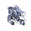
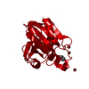
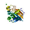
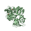
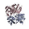
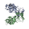
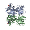
| Entry | Database: PDB / ID: 2vl9 | ||||||
|---|---|---|---|---|---|---|---|
| Title | Oxidized form of human peroxiredoxin 5 | ||||||
 Components Components | PEROXIREDOXIN-5 | ||||||
 Keywords Keywords | OXIDOREDUCTASE / THIOREDOXIN PEROXIDASE / ALTERNATIVE INITIATION / ANTIOXIDANT ENZYME / REDOX-ACTIVE CENTER / CYTOPLASM / PEROXIDASE / PEROXISOME / ANTIOXIDANT / POLYMORPHISM / MITOCHONDRION / PEROXIREDOXIN / TRANSIT PEPTIDE / THIOREDOXIN FOLD | ||||||
| Function / homology |  Function and homology information Function and homology informationperoxynitrite reductase activity / reactive nitrogen species metabolic process / negative regulation of transcription by RNA polymerase III / negative regulation of oxidoreductase activity / NADPH oxidation / regulation of apoptosis involved in tissue homeostasis / RNA polymerase III transcription regulatory region sequence-specific DNA binding / thioredoxin-dependent peroxiredoxin / thioredoxin peroxidase activity / Detoxification of Reactive Oxygen Species ...peroxynitrite reductase activity / reactive nitrogen species metabolic process / negative regulation of transcription by RNA polymerase III / negative regulation of oxidoreductase activity / NADPH oxidation / regulation of apoptosis involved in tissue homeostasis / RNA polymerase III transcription regulatory region sequence-specific DNA binding / thioredoxin-dependent peroxiredoxin / thioredoxin peroxidase activity / Detoxification of Reactive Oxygen Species / antioxidant activity / cysteine-type endopeptidase inhibitor activity involved in apoptotic process / peroxisomal matrix / positive regulation of collagen biosynthetic process / cell redox homeostasis / hydrogen peroxide catabolic process / TP53 Regulates Metabolic Genes / peroxidase activity / cellular response to reactive oxygen species / peroxisome / cellular response to oxidative stress / cytoplasmic vesicle / response to oxidative stress / mitochondrial matrix / inflammatory response / signaling receptor binding / intracellular membrane-bounded organelle / negative regulation of apoptotic process / perinuclear region of cytoplasm / mitochondrion / extracellular space / extracellular exosome / identical protein binding / nucleus / cytosol / cytoplasmSimilarity search - Function | ||||||
| Biological species |  HOMO SAPIENS (human) HOMO SAPIENS (human) | ||||||
| Method | X-RAY DIFFRACTION / SYNCHROTRON / MOLECULAR REPLACEMENT / Resolution: 2.7 Å | ||||||
 Authors Authors | Smeets, A. / Declercq, J.P. | ||||||
 Citation Citation | Journal: Arch.Biochem.Biophys. / Year: 2008 Title: The Crystal Structures of Oxidized Forms of Human Peroxiredoxin 5 with an Intramolecular Disulfide Bond Confirm the Proposed Enzymatic Mechanism for Atypical 2-Cys Peroxiredoxins. Authors: Smeets, A. / Marchand, C. / Linard, D. / Knoops, B. / Declercq, J.P. | ||||||
| History |
|
- Structure visualization
| Structure viewer | Molecule: MolmilJmol/JSmol |
|---|
- Downloads & links
-Download
| PDBx/mmCIF format | 2vl9.cif.gz | 129.5 KB | Display | PDBx/mmCIF format |
|---|---|---|---|---|
| PDB format | pdb2vl9.ent.gz | 101.6 KB | Display | PDB format |
| PDBx/mmJSON format | 2vl9.json.gz | Tree view | PDBx/mmJSON format | |
| Others |  Other downloads Other downloads |
-Validation report
| Arichive directory | https://data.pdbj.org/pub/pdb/validation_reports/vl/2vl9ftp://data.pdbj.org/pub/pdb/validation_reports/vl/2vl9 | HTTPS FTP |
|---|
-Related structure data
| Related structure data | 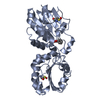 2vl2C 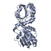 2vl3C 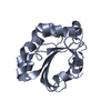 1oc3S S: Starting model for refinement C: citing same article ( |
|---|---|
| Similar structure data |


-Links
 PDBj
PDBj


- Assembly
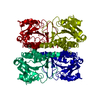
| Deposited unit | 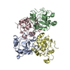
| ||||||||
|---|---|---|---|---|---|---|---|---|---|
| 1 |
| ||||||||
| Unit cell |
|
-Components
| #1: Protein | / PRX-V / PEROXISOMAL ANTIOXIDANT ENZYME / PLP / THIOREDOXIN REDUCTASE / THIOREDOXIN PEROXIDASE PMP20 ...PRX-V / PEROXISOMAL ANTIOXIDANT ENZYME / PLP / THIOREDOXIN REDUCTASE / THIOREDOXIN PEROXIDASE PMP20 / ANTIOXIDANT ENZYME B166 / AOEB166 / TPX TYPE VI / LIVER TISSUE 2D-PAGE SPOT 71B / ALU COREPRESSOR 1 / HUMAN PEROXIREDOXIN 5 Mass: 18308.979 Da / Num. of mol.: 4 / Fragment: RESIDUES 2-162 / Mutation: YES Source method: isolated from a genetically manipulated source Source: (gene. exp.) HOMO SAPIENS (human) / Organ: LUNG / Plasmid: PQE-30 / Production host:  ESCHERICHIA COLI M15 (bacteria) / References: UniProt: P30044, peroxiredoxin ESCHERICHIA COLI M15 (bacteria) / References: UniProt: P30044, peroxiredoxin#2: Water | ChemComp-HOH / | Water Mass: 18.015 Da / Num. of mol.: 57 / Source method: isolated from a natural source / Formula: H2O Mass: 18.015 Da / Num. of mol.: 57 / Source method: isolated from a natural source / Formula: H2OCompound details | ENGINEERED RESIDUE IN CHAIN A, CYS 73 TO SER ENGINEERED RESIDUE IN CHAIN B, CYS 73 TO SER ...ENGINEERED | |
|---|
-Experimental details
-Experiment
| Experiment | Method: X-RAY DIFFRACTION / Number of used crystals: 1 |
|---|
- Sample preparation
| Crystal | Density Matthews: 2.22 Å3/Da / Density % sol: 44.53 % Description: THE MODEL USED FOR THE MOLECULAR REPLACEMENT WAS A SIMULATION CREATED FROM PDB ENTRY 1OC3 |
|---|---|
| Crystal grow | pH: 6.5 Details: PEG 8000 20% SODIUM CACODYLATE 0.1 M PH 6.5 SODIUM ACETATE 0.1 M 6-AMINOCAPROIC ACID 3% W/V |
-Data collection
| Diffraction | Mean temperature: 100 K |
|---|---|
| Diffraction source | Source: SYNCHROTRON / Site: ESRF  / Beamline: BM30A / Wavelength: 0.979764 / Beamline: BM30A / Wavelength: 0.979764 |
| Detector | Type: MARRESEARCH / Detector: CCD / Date: Nov 11, 2005 / Details: TWO MIRRORS ARE USED FOR VERTICAL FOCUSSING |
| Radiation | Monochromator: FIRST CRYSTAL FLAT AND N2 COOLED. SECOND ONE SAGITALLY BENT Protocol: SINGLE WAVELENGTH / Monochromatic (M) / Laue (L): M / Scattering type: x-ray |
| Radiation wavelength | Wavelength: 0.979764 Å / Relative weight: 1 |
| Reflection twin | Operator: -K,H+K,L / Fraction: 0.479 |
| Reflection | Resolution: 2.7→46 Å / Num. obs: 18076 / % possible obs: 98.1 % / Observed criterion σ(I): 0 / Redundancy: 4.05 % / Rmerge(I) obs: 0.05 / Net I/σ(I): 17.9 |
| Reflection shell | Resolution: 2.7→2.75 Å / Redundancy: 4.05 % / Rmerge(I) obs: 0.46 / Mean I/σ(I) obs: 3.4 / % possible all: 99.6 |
- Processing
| Software |
| ||||||||||||||||||||||||
|---|---|---|---|---|---|---|---|---|---|---|---|---|---|---|---|---|---|---|---|---|---|---|---|---|---|
| Refinement | Method to determine structure: MOLECULAR REPLACEMENT Starting model: PDB ENTRY 1OC3 Resolution: 2.7→46 Å / Phase error: 24.5 / Stereochemistry target values: TWIN_LSQ_F Details: TWIN RATIO IS 50%. COMPUTED STRUCTURE FACTORS ARE NOT DIRECTLY COMPARABLE TO THE DEPOSITED OBSERVED STRUCTURE FACTORS.
| ||||||||||||||||||||||||
| Solvent computation | Shrinkage radii: 0.9 Å / VDW probe radii: 1.11 Å / Solvent model: FLAT BULK SOLVENT MODEL / Bsol: 170.46 Å2 / ksol: 0.37 e/Å3 | ||||||||||||||||||||||||
| Displacement parameters | Biso mean: 79.53 Å2
| ||||||||||||||||||||||||
| Refinement step | Cycle: LAST / Resolution: 2.7→46 Å
| ||||||||||||||||||||||||
| Refine LS restraints |
|
