 Movie
Movie Controller

[English] 日本語
 Yorodumi
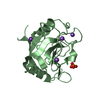
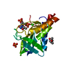
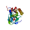
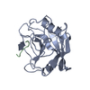
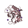
- PDB-5wsz: Crystal structure of a lytic polysaccharide monooxygenase,BtLPMO1... -
+ Open data

- Basic information
| Entry | Database: PDB / ID: 5wsz | ||||||
|---|---|---|---|---|---|---|---|
| Title | Crystal structure of a lytic polysaccharide monooxygenase,BtLPMO10A, from Bacillus thuringiensis | ||||||
 Components Components | LpmO10A | ||||||
 Keywords Keywords |  OXIDOREDUCTASE / copper-dependent / monooxygenase / AA10 OXIDOREDUCTASE / copper-dependent / monooxygenase / AA10 | ||||||
| Function / homology |  Function and homology information Function and homology information | ||||||
| Biological species |  Bacillus thuringiensis subsp. kurstaki (bacteria) Bacillus thuringiensis subsp. kurstaki (bacteria) | ||||||
| Method | X-RAY DIFFRACTION / SYNCHROTRON / MOLECULAR REPLACEMENT / Resolution: 2.565 Å | ||||||
 Authors Authors | Zhao, Y. / Zhang, H. / Yin, H. | ||||||
| Funding support |  China, 1items China, 1items
| ||||||
 Citation Citation | Journal: To Be Published Title: Crystal structure of a lytic polysaccharide monooxygenase,BtLPMO10A, from Bacillus thuringiensis Authors: Zhao, Y. / Zhang, H. / Yin, H. | ||||||
| History |
|
- Structure visualization
| Structure viewer | Molecule: MolmilJmol/JSmol |
|---|
- Downloads & links
-Download
| PDBx/mmCIF format | 5wsz.cif.gz | 141.7 KB | Display | PDBx/mmCIF format |
|---|---|---|---|---|
| PDB format | pdb5wsz.ent.gz | 110.7 KB | Display | PDB format |
| PDBx/mmJSON format | 5wsz.json.gz | Tree view | PDBx/mmJSON format | |
| Others |  Other downloads Other downloads |
-Validation report
| Arichive directory | https://data.pdbj.org/pub/pdb/validation_reports/ws/5wszftp://data.pdbj.org/pub/pdb/validation_reports/ws/5wsz | HTTPS FTP |
|---|
-Related structure data
| Related structure data | 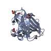 2bemS S: Starting model for refinement |
|---|---|
| Similar structure data |


-Links
 PDBj
PDBj- Assembly
| Deposited unit | 
| ||||||||
|---|---|---|---|---|---|---|---|---|---|
| 1 | 
| ||||||||
| 2 | 
| ||||||||
| 3 | 
| ||||||||
| 4 | 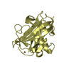
| ||||||||
| Unit cell |
|
-Components
| #1: Protein | Mass: 18622.801 Da / Num. of mol.: 4 / Fragment: UNP residues 35-203 Source method: isolated from a genetically manipulated source Source: (gene. exp.) Bacillus thuringiensis subsp. kurstaki (bacteria)Gene: lpmO10A / Production host: Escherichia coli BL21(DE3) (bacteria) / Strain (production host): BL21(DE3) / References: UniProt: A0A0C5K362, UniProt: A0A0F6FRT6*PLUS#2: Chemical | ChemComp-CU / Copper  Mass: 63.546 Da / Num. of mol.: 4 / Source method: obtained synthetically / Formula: Cu Mass: 63.546 Da / Num. of mol.: 4 / Source method: obtained synthetically / Formula: Cu#3: Water | ChemComp-HOH / | Water Mass: 18.015 Da / Num. of mol.: 130 / Source method: isolated from a natural source / Formula: H2O Mass: 18.015 Da / Num. of mol.: 130 / Source method: isolated from a natural source / Formula: H2O |
|---|
-Experimental details
-Experiment
| Experiment | Method: X-RAY DIFFRACTION / Number of used crystals: 1 |
|---|
- Sample preparation
| Crystal | Density Matthews: 2.05 Å3/Da / Density % sol: 40.11 % |
|---|---|
| Crystal grow | Temperature: 293 K / Method: vapor diffusion, sitting drop / pH: 6.5 Details: 0.2M magnesium acetate tetrahydrate, 0.1M sodium cacodylate trihydrate pH 6.5, 20% w/v Polyethylene glycol 8000 |
-Data collection
| Diffraction | Mean temperature: 100 K |
|---|---|
| Diffraction source | Source: SYNCHROTRON / Site: SSRF / Beamline: BL18U1 / Wavelength: 0.97776 Å |
| Detector | Type: MARMOSAIC 325 mm CCD / Detector: CCD / Date: Oct 17, 2016 |
| Radiation | Protocol: SINGLE WAVELENGTH / Monochromatic (M) / Laue (L): M / Scattering type: x-ray |
| Radiation wavelength | Wavelength: 0.97776 Å / Relative weight: 1 |
| Reflection | Resolution: 2.57→50 Å / Num. obs: 20252 / % possible obs: 100 % / Redundancy: 7.5 % / Rmerge(I) obs: 0.195 / Rpim(I) all: 0.072 / Net I/σ(I): 10.9 |
| Reflection shell | Resolution: 2.57→2.66 Å / Rmerge(I) obs: 0.784 / Rpim(I) all: 0.31 / % possible all: 91.06 |
- Processing
| Software |
| |||||||||||||||||||||||||||||||||||||||||||||||||
|---|---|---|---|---|---|---|---|---|---|---|---|---|---|---|---|---|---|---|---|---|---|---|---|---|---|---|---|---|---|---|---|---|---|---|---|---|---|---|---|---|---|---|---|---|---|---|---|---|---|---|
| Refinement | Method to determine structure: MOLECULAR REPLACEMENT Starting model: 2BEM Resolution: 2.565→47.118 Å / SU ML: 0.27 / Cross valid method: THROUGHOUT / σ(F): 1.34 / Phase error: 28.7 / Stereochemistry target values: ML
| |||||||||||||||||||||||||||||||||||||||||||||||||
| Solvent computation | Shrinkage radii: 0.9 Å / VDW probe radii: 1.11 Å / Solvent model: FLAT BULK SOLVENT MODEL | |||||||||||||||||||||||||||||||||||||||||||||||||
| Refinement step | Cycle: LAST / Resolution: 2.565→47.118 Å
| |||||||||||||||||||||||||||||||||||||||||||||||||
| Refine LS restraints |
| |||||||||||||||||||||||||||||||||||||||||||||||||
| LS refinement shell |
|
