 Movie
Movie Controller

[English] 日本語
 Yorodumi
- PDB-4h87: Crystal structure of a FHA domain of kanadaptin (SLC4A1AP) from H... -
+ Open data

- Basic information
| Entry | Database: PDB / ID: 4h87 | ||||||
|---|---|---|---|---|---|---|---|
| Title | Crystal structure of a FHA domain of kanadaptin (SLC4A1AP) from Homo sapiens at 1.55 A resolution | ||||||
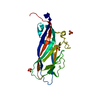
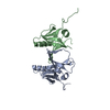
 Components Components | Kanadaptin | ||||||
 Keywords Keywords | PEPTIDE BINDING PROTEIN / FHA domain of PF00498 /  mRNA processing / nucleus / Structural Genomics / Joint Center for Structural Genomics / JCSG / Protein Structure Initiative / PSI-BIOLOGY / SIGNAL TRANSDUCTION mRNA processing / nucleus / Structural Genomics / Joint Center for Structural Genomics / JCSG / Protein Structure Initiative / PSI-BIOLOGY / SIGNAL TRANSDUCTION | ||||||
| Function / homology |  Function and homology information Function and homology informationintracellular membrane-bounded organelle / mRNA binding / nucleoplasm / plasma membrane / cytoplasmSimilarity search - Function | ||||||
| Biological species |  Homo sapiens (human) Homo sapiens (human) | ||||||
| Method | X-RAY DIFFRACTION / SYNCHROTRON / MOLECULAR REPLACEMENT / molecular replacement / Resolution: 1.55 Å | ||||||
 Authors Authors | Joint Center for Structural Genomics (JCSG) | ||||||
 Citation Citation | Journal: To be published Title: Crystal structure of a FHA domain of kanadaptin (SLC4A1AP) from Homo sapiens at 1.55 A resolution Authors: Joint Center for Structural Genomics (JCSG) | ||||||
| History |
|
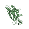
- Structure visualization
| Structure viewer | Molecule: MolmilJmol/JSmol |
|---|
- Downloads & links
-Download
| PDBx/mmCIF format | 4h87.cif.gz | 118.1 KB | Display | PDBx/mmCIF format |
|---|---|---|---|---|
| PDB format | pdb4h87.ent.gz | 91.4 KB | Display | PDB format |
| PDBx/mmJSON format | 4h87.json.gz | Tree view | PDBx/mmJSON format | |
| Others |  Other downloads Other downloads |
-Validation report
| Arichive directory | https://data.pdbj.org/pub/pdb/validation_reports/h8/4h87ftp://data.pdbj.org/pub/pdb/validation_reports/h8/4h87 | HTTPS FTP |
|---|
-Related structure data
| Related structure data |  3elsS S: Starting model for refinement |
|---|---|
| Similar structure data | |
| Other databases |


-Links
 PDBj
PDBj

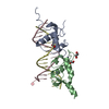
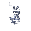
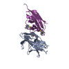
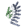
- Assembly
| Deposited unit | 
| ||||||||
|---|---|---|---|---|---|---|---|---|---|
| 1 |
| ||||||||
| 2 | 
| ||||||||
| 3 | 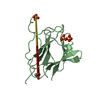
| ||||||||
| Unit cell |
|
-Components
| #1: Protein | Mass: 14101.977 Da / Num. of mol.: 2 / Fragment: FHA domain Source method: isolated from a genetically manipulated source Source: (gene. exp.) Homo sapiens (human) / Gene: HLC3, NM_018158, SLC4A1AP / Plasmid: SGC_M / Production host:  Escherichia Coli (E. coli) / Strain (production host): Rosetta / References: UniProt: Q9BWU0 Escherichia Coli (E. coli) / Strain (production host): Rosetta / References: UniProt: Q9BWU0#2: Chemical | ChemComp-SO4 / Sulfate  Mass: 96.063 Da / Num. of mol.: 6 / Source method: obtained synthetically / Formula: SO4 Mass: 96.063 Da / Num. of mol.: 6 / Source method: obtained synthetically / Formula: SO4#3: Chemical | ChemComp-GOL / Glycerol  Mass: 92.094 Da / Num. of mol.: 5 / Source method: obtained synthetically / Formula: C3H8O3 Mass: 92.094 Da / Num. of mol.: 5 / Source method: obtained synthetically / Formula: C3H8O3#4: Water | ChemComp-HOH / | Water Mass: 18.015 Da / Num. of mol.: 207 / Source method: isolated from a natural source / Formula: H2O Mass: 18.015 Da / Num. of mol.: 207 / Source method: isolated from a natural source / Formula: H2OSequence details | THE CONSTRUCT WAS EXPRESSED WITH AN N-TERMINAL PURIFICATION TAG CONTAINING 6HIS-HA-FLAG-TEV SITES - ...THE CONSTRUCT WAS EXPRESSED WITH AN N-TERMINAL PURIFICATI | |
|---|
-Experimental details
-Experiment
| Experiment | Method: X-RAY DIFFRACTION / Number of used crystals: 1 |
|---|
- Sample preparation
| Crystal | Density Matthews: 2.09 Å3/Da / Density % sol: 41.08 % Description: THE INPUT MODEL WAS PREPARED USING PHENIX.ROSETTA FOLLOWING THE PROCEDURE USED IN THE JCSG MR-PIPELINE (PMID: 18094477) |
|---|---|
| Crystal grow | Temperature: 277 K / Method: vapor diffusion, sitting drop / pH: 5 Details: 1.60M ammonium sulfate, 0.1M citric acid pH 5.0, NANODROP, VAPOR DIFFUSION, SITTING DROP, temperature 277K |
-Data collection
| Diffraction | Mean temperature: 100 K | |||||||||||||||||||||||||||||||||||||||||||||||||||||||||||||||||||||||||||||
|---|---|---|---|---|---|---|---|---|---|---|---|---|---|---|---|---|---|---|---|---|---|---|---|---|---|---|---|---|---|---|---|---|---|---|---|---|---|---|---|---|---|---|---|---|---|---|---|---|---|---|---|---|---|---|---|---|---|---|---|---|---|---|---|---|---|---|---|---|---|---|---|---|---|---|---|---|---|---|
| Diffraction source | Source: SYNCHROTRON / Site: SSRL  / Beamline: BL11-1 / Wavelength: 0.97932 / Beamline: BL11-1 / Wavelength: 0.97932 | |||||||||||||||||||||||||||||||||||||||||||||||||||||||||||||||||||||||||||||
| Detector | Type: DECTRIS PILATUS 6M / Detector: PIXEL / Date: Aug 1, 2012 Details: Flat mirror (vertical focusing); single crystal Si(111) bent monochromator (horizontal focusing) | |||||||||||||||||||||||||||||||||||||||||||||||||||||||||||||||||||||||||||||
| Radiation | Monochromator: single crystal Si(111) bent / Protocol: SINGLE WAVELENGTH / Monochromatic (M) / Laue (L): M / Scattering type: x-ray | |||||||||||||||||||||||||||||||||||||||||||||||||||||||||||||||||||||||||||||
| Radiation wavelength | Wavelength: 0.97932 Å / Relative weight: 1 | |||||||||||||||||||||||||||||||||||||||||||||||||||||||||||||||||||||||||||||
| Reflection | Resolution: 1.55→36.764 Å / Num. obs: 35157 / % possible obs: 99.9 % / Observed criterion σ(I): -3 / Biso Wilson estimate: 20.678 Å2 / Rmerge(I) obs: 0.048 / Net I/σ(I): 27.74 | |||||||||||||||||||||||||||||||||||||||||||||||||||||||||||||||||||||||||||||
| Reflection shell |
|
-Phasing
| Phasing | Method: molecular replacement |
|---|
- Processing
| Software |
| ||||||||||||||||||||||||||||||||||||||||||||||||||||||||||||||||||||||||||||||||||||||||||||||||||||||||||||
|---|---|---|---|---|---|---|---|---|---|---|---|---|---|---|---|---|---|---|---|---|---|---|---|---|---|---|---|---|---|---|---|---|---|---|---|---|---|---|---|---|---|---|---|---|---|---|---|---|---|---|---|---|---|---|---|---|---|---|---|---|---|---|---|---|---|---|---|---|---|---|---|---|---|---|---|---|---|---|---|---|---|---|---|---|---|---|---|---|---|---|---|---|---|---|---|---|---|---|---|---|---|---|---|---|---|---|---|---|---|
| Refinement | Method to determine structure: MOLECULAR REPLACEMENT Starting model: PDB entry 3ELS Resolution: 1.55→36.764 Å / Cor.coef. Fo:Fc: 0.961 / Cor.coef. Fo:Fc free: 0.9492 / Occupancy max: 1 / Occupancy min: 0.3 / Cross valid method: THROUGHOUT / σ(F): 0 Details: 1. ATOM RECORD CONTAINS SUM OF TLS AND RESIDUAL B FACTORS. ANISOU RECORD CONTAINS SUM OF TLS AND RESIDUAL U FACTORS. 2. SO4 AND GOL MODELED ARE PRESENT IN CRYSTALLIZATION/CRYO CONDITIONS. 3. ...Details: 1. ATOM RECORD CONTAINS SUM OF TLS AND RESIDUAL B FACTORS. ANISOU RECORD CONTAINS SUM OF TLS AND RESIDUAL U FACTORS. 2. SO4 AND GOL MODELED ARE PRESENT IN CRYSTALLIZATION/CRYO CONDITIONS. 3. NCS RESTRAINTS WERE APPLIED USING BUSTER'S LSSR RESTRAINT REPRESENTATION (-AUTONCS)
| ||||||||||||||||||||||||||||||||||||||||||||||||||||||||||||||||||||||||||||||||||||||||||||||||||||||||||||
| Displacement parameters | Biso max: 104.2 Å2 / Biso mean: 26.6599 Å2 / Biso min: 11.21 Å2
| ||||||||||||||||||||||||||||||||||||||||||||||||||||||||||||||||||||||||||||||||||||||||||||||||||||||||||||
| Refine analyze | Luzzati coordinate error obs: 0.183 Å | ||||||||||||||||||||||||||||||||||||||||||||||||||||||||||||||||||||||||||||||||||||||||||||||||||||||||||||
| Refinement step | Cycle: LAST / Resolution: 1.55→36.764 Å
| ||||||||||||||||||||||||||||||||||||||||||||||||||||||||||||||||||||||||||||||||||||||||||||||||||||||||||||
| Refine LS restraints |
| ||||||||||||||||||||||||||||||||||||||||||||||||||||||||||||||||||||||||||||||||||||||||||||||||||||||||||||
| LS refinement shell | Resolution: 1.55→1.59 Å / Total num. of bins used: 18
| ||||||||||||||||||||||||||||||||||||||||||||||||||||||||||||||||||||||||||||||||||||||||||||||||||||||||||||
| Refinement TLS params. | Method: refined / Refine-ID: X-RAY DIFFRACTION
| ||||||||||||||||||||||||||||||||||||||||||||||||||||||||||||||||||||||||||||||||||||||||||||||||||||||||||||
| Refinement TLS group |
|
