 Movie
Movie Controller

[English] 日本語
 Yorodumi
- PDB-2d2a: Crystal Structure of Escherichia coli SufA Involved in Biosynthes... -
+ Open data

- Basic information
| Entry | Database: PDB / ID: 2d2a | ||||||
|---|---|---|---|---|---|---|---|
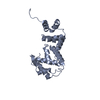
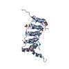
| Title | Crystal Structure of Escherichia coli SufA Involved in Biosynthesis of Iron-sulfur Clusters | ||||||
 Components Components | SufA protein | ||||||
 Keywords Keywords | METAL TRANSPORT /  Iron-sulfur cluster / iron / SUF / SufA / IscA / YadR Iron-sulfur cluster / iron / SUF / SufA / IscA / YadR | ||||||
| Function / homology |  Function and homology information Function and homology informationprotein maturation by iron-sulfur cluster transfer / iron-sulfur cluster assembly / 2 iron, 2 sulfur cluster binding / response to oxidative stress / structural molecule activity / cytosol / cytoplasmSimilarity search - Function | ||||||
| Biological species |  Escherichia coli (E. coli) Escherichia coli (E. coli) | ||||||
| Method | X-RAY DIFFRACTION / SYNCHROTRON / MOLECULAR REPLACEMENT / Resolution: 2.7 Å | ||||||
 Authors Authors | Wada, K. / Hasegawa, Y. / Gong, Z. / Minami, Y. / Fukuyama, K. / Takahashi, Y. | ||||||
 Citation Citation | Journal: Febs Lett. / Year: 2005 Title: Crystal structure of Escherichia coli SufA involved in biosynthesis of iron-sulfur clusters: Implications for a functional dimer Authors: Wada, K. / Hasegawa, Y. / Gong, Z. / Minami, Y. / Fukuyama, K. / Takahashi, Y. | ||||||
| History |
|
- Structure visualization
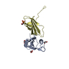
| Structure viewer | Molecule: MolmilJmol/JSmol |
|---|
- Downloads & links
-Download
| PDBx/mmCIF format | 2d2a.cif.gz | 57.4 KB | Display | PDBx/mmCIF format |
|---|---|---|---|---|
| PDB format | pdb2d2a.ent.gz | 40.4 KB | Display | PDB format |
| PDBx/mmJSON format | 2d2a.json.gz | Tree view | PDBx/mmJSON format | |
| Others |  Other downloads Other downloads |
-Validation report
| Arichive directory | https://data.pdbj.org/pub/pdb/validation_reports/d2/2d2aftp://data.pdbj.org/pub/pdb/validation_reports/d2/2d2a | HTTPS FTP |
|---|
-Related structure data
| Related structure data | 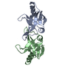 1s98S S: Starting model for refinement |
|---|---|
| Similar structure data |


-Links
 PDBj
PDBj- Assembly
| Deposited unit | 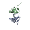
| ||||||||
|---|---|---|---|---|---|---|---|---|---|
| 1 |
| ||||||||
| 2 | 
| ||||||||
| Unit cell |
| ||||||||
| Details | The biological assembly is a dimer in the asymmetric unit. |
-Components
| #1: Protein | Mass: 16076.958 Da / Num. of mol.: 2 Source method: isolated from a genetically manipulated source Source: (gene. exp.) Escherichia coli (E. coli) / Plasmid: pET-19b / Production host: Escherichia coli (E. coli) / Strain (production host): C41(DE3) / References: UniProt: P77667#2: Water | ChemComp-HOH / | Water Mass: 18.015 Da / Num. of mol.: 37 / Source method: isolated from a natural source / Formula: H2O Mass: 18.015 Da / Num. of mol.: 37 / Source method: isolated from a natural source / Formula: H2O |
|---|
-Experimental details
-Experiment
| Experiment | Method: X-RAY DIFFRACTION / Number of used crystals: 1 |
|---|
- Sample preparation
| Crystal | Density Matthews: 2.1 Å3/Da / Density % sol: 41.2 % |
|---|---|
| Crystal grow | Temperature: 293 K / Method: vapor diffusion, hanging drop / pH: 4.8 Details: PEG2000MME, pH 4.8, VAPOR DIFFUSION, HANGING DROP, temperature 293K |
-Data collection
| Diffraction | Mean temperature: 100 K |
|---|---|
| Diffraction source | Source: SYNCHROTRON / Site: SPring-8  / Beamline: BL41XU / Wavelength: 1 Å / Beamline: BL41XU / Wavelength: 1 Å |
| Detector | Type: MARRESEARCH / Detector: CCD / Date: May 29, 2004 |
| Radiation | Monochromator: SI(111) DOUBLE MONOCHROMATOR / Protocol: SINGLE WAVELENGTH / Monochromatic (M) / Laue (L): M / Scattering type: x-ray |
| Radiation wavelength | Wavelength: 1 Å / Relative weight: 1 |
| Reflection | Resolution: 2.7→50 Å / Num. obs: 8136 / % possible obs: 99.9 % / Observed criterion σ(F): 1 / Observed criterion σ(I): 1 / Biso Wilson estimate: 40.1 Å2 / Rmerge(I) obs: 0.085 |
| Reflection shell | Resolution: 2.7→2.8 Å / Redundancy: 6.8 % / Rmerge(I) obs: 0.349 / Num. unique all: 808 / % possible all: 100 |
- Processing
| Software |
| |||||||||||||||||||||||||
|---|---|---|---|---|---|---|---|---|---|---|---|---|---|---|---|---|---|---|---|---|---|---|---|---|---|---|
| Refinement | Method to determine structure: MOLECULAR REPLACEMENT Starting model: PDB ENTRY 1S98 Resolution: 2.7→41.6 Å / Rfactor Rfree error: 0.01 / Data cutoff high absF: 421277.64 / Data cutoff low absF: 0 / Isotropic thermal model: RESTRAINED / Cross valid method: THROUGHOUT / σ(F): 0 / Stereochemistry target values: Engh & Huber
| |||||||||||||||||||||||||
| Solvent computation | Solvent model: FLAT MODEL / Bsol: 43.8321 Å2 / ksol: 0.370499 e/Å3 | |||||||||||||||||||||||||
| Displacement parameters | Biso mean: 34.9 Å2
| |||||||||||||||||||||||||
| Refine analyze |
| |||||||||||||||||||||||||
| Refinement step | Cycle: LAST / Resolution: 2.7→41.6 Å
| |||||||||||||||||||||||||
| Refine LS restraints |
| |||||||||||||||||||||||||
| LS refinement shell | Resolution: 2.7→2.87 Å / Rfactor Rfree error: 0.027 / Total num. of bins used: 6
| |||||||||||||||||||||||||
| Xplor file |
|
