 Movie
Movie Controller


+ Open data

- Basic information
| Entry | Database: PDB / ID: 3t04 | ||||||
|---|---|---|---|---|---|---|---|
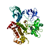
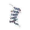
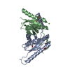
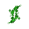
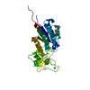
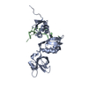
| Title | Crystal structure of monobody 7c12/abl1 sh2 domain complex | ||||||
 Components Components |
| ||||||
 Keywords Keywords |  SIGNALING PROTEIN/PROTEIN BINDING / ENGINEERED BINDING PROTEIN / ANTIBODY MIMIC / PROTEIN-PROTEIN COMPLEX / SH2 DOMAIN / ATP-BINDING / PHOSPHOPROTEIN / TYROSINE-PROTEIN KINASE / SIGNALING PROTEIN-PROTEIN BINDING complex SIGNALING PROTEIN/PROTEIN BINDING / ENGINEERED BINDING PROTEIN / ANTIBODY MIMIC / PROTEIN-PROTEIN COMPLEX / SH2 DOMAIN / ATP-BINDING / PHOSPHOPROTEIN / TYROSINE-PROTEIN KINASE / SIGNALING PROTEIN-PROTEIN BINDING complex | ||||||
| Function / homology |  Function and homology information: / positive regulation of actin filament binding / positive regulation of oxidoreductase activity / protein localization to cytoplasmic microtubule plus-end / DNA conformation change / podocyte apoptotic process / DN4 thymocyte differentiation / Role of ABL in ROBO-SLIT signaling / response to epinephrine / transitional one stage B cell differentiation ...: / positive regulation of actin filament binding / positive regulation of oxidoreductase activity / protein localization to cytoplasmic microtubule plus-end / DNA conformation change / podocyte apoptotic process / DN4 thymocyte differentiation / Role of ABL in ROBO-SLIT signaling / response to epinephrine / transitional one stage B cell differentiation / activation of protein kinase C activity / nicotinate-nucleotide adenylyltransferase activity / regulation of modification of synaptic structure / positive regulation of microtubule binding / delta-catenin binding / B cell proliferation involved in immune response / positive regulation of extracellular matrix organization / neuroepithelial cell differentiation / microspike assembly / positive regulation of Wnt signaling pathway, planar cell polarity pathway / cerebellum morphogenesis / positive regulation of blood vessel branching / B-1 B cell homeostasis / mitochondrial depolarization / negative regulation of ubiquitin-protein transferase activity / neuropilin signaling pathway / neuropilin binding / bubble DNA binding / negative regulation of protein serine/threonine kinase activity / activated T cell proliferation / cellular response to dopamine / regulation of cell motility / regulation of Cdc42 protein signal transduction / proline-rich region binding / positive regulation of dendrite development / mitogen-activated protein kinase binding / myoblast proliferation / alpha-beta T cell differentiation / regulation of hematopoietic stem cell differentiation / syntaxin binding / cardiac muscle cell proliferation / regulation of T cell differentiation / regulation of axon extension / HDR through Single Strand Annealing (SSA) / positive regulation of cell migration involved in sprouting angiogenesis / negative regulation of cell-cell adhesion / Fc-gamma receptor signaling pathway involved in phagocytosis / Myogenesis / regulation of microtubule polymerization / positive regulation of osteoblast proliferation / RUNX2 regulates osteoblast differentiation / platelet-derived growth factor receptor-beta signaling pathway / positive regulation of focal adhesion assembly / negative regulation of cellular senescence / Bergmann glial cell differentiation / associative learning / neuromuscular process controlling balance / regulation of endocytosis / negative regulation of BMP signaling pathway / negative regulation of mitotic cell cycle / actin monomer binding / negative regulation of long-term synaptic potentiation / endothelial cell migration / RHO GTPases Activate WASPs and WAVEs / positive regulation of T cell migration / canonical NF-kappaB signal transduction / signal transduction in response to DNA damage / negative regulation of double-strand break repair via homologous recombination / mismatch repair / BMP signaling pathway / regulation of cell adhesion / negative regulation of endothelial cell apoptotic process / four-way junction DNA binding / positive regulation of substrate adhesion-dependent cell spreading / peptidyl-tyrosine autophosphorylation / positive regulation of vasoconstriction / positive regulation of stress fiber assembly / spleen development / ruffle / cellular response to transforming growth factor beta stimulus / positive regulation of establishment of T cell polarity / ERK1 and ERK2 cascade / positive regulation of interleukin-2 production / actin filament polymerization / phosphotyrosine residue binding / SH2 domain binding / response to endoplasmic reticulum stress / ephrin receptor binding / positive regulation of endothelial cell migration / positive regulation of mitotic cell cycle / substrate adhesion-dependent cell spreading / positive regulation of release of sequestered calcium ion into cytosol / post-embryonic development / thymus development / regulation of autophagy / neural tube closure / establishment of localization in cell / integrin-mediated signaling pathway / regulation of actin cytoskeleton organization / protein kinase C binding Function and homology information: / positive regulation of actin filament binding / positive regulation of oxidoreductase activity / protein localization to cytoplasmic microtubule plus-end / DNA conformation change / podocyte apoptotic process / DN4 thymocyte differentiation / Role of ABL in ROBO-SLIT signaling / response to epinephrine / transitional one stage B cell differentiation ...: / positive regulation of actin filament binding / positive regulation of oxidoreductase activity / protein localization to cytoplasmic microtubule plus-end / DNA conformation change / podocyte apoptotic process / DN4 thymocyte differentiation / Role of ABL in ROBO-SLIT signaling / response to epinephrine / transitional one stage B cell differentiation / activation of protein kinase C activity / nicotinate-nucleotide adenylyltransferase activity / regulation of modification of synaptic structure / positive regulation of microtubule binding / delta-catenin binding / B cell proliferation involved in immune response / positive regulation of extracellular matrix organization / neuroepithelial cell differentiation / microspike assembly / positive regulation of Wnt signaling pathway, planar cell polarity pathway / cerebellum morphogenesis / positive regulation of blood vessel branching / B-1 B cell homeostasis / mitochondrial depolarization / negative regulation of ubiquitin-protein transferase activity / neuropilin signaling pathway / neuropilin binding / bubble DNA binding / negative regulation of protein serine/threonine kinase activity / activated T cell proliferation / cellular response to dopamine / regulation of cell motility / regulation of Cdc42 protein signal transduction / proline-rich region binding / positive regulation of dendrite development / mitogen-activated protein kinase binding / myoblast proliferation / alpha-beta T cell differentiation / regulation of hematopoietic stem cell differentiation / syntaxin binding / cardiac muscle cell proliferation / regulation of T cell differentiation / regulation of axon extension / HDR through Single Strand Annealing (SSA) / positive regulation of cell migration involved in sprouting angiogenesis / negative regulation of cell-cell adhesion / Fc-gamma receptor signaling pathway involved in phagocytosis / Myogenesis / regulation of microtubule polymerization / positive regulation of osteoblast proliferation / RUNX2 regulates osteoblast differentiation / platelet-derived growth factor receptor-beta signaling pathway / positive regulation of focal adhesion assembly / negative regulation of cellular senescence / Bergmann glial cell differentiation / associative learning / neuromuscular process controlling balance / regulation of endocytosis / negative regulation of BMP signaling pathway / negative regulation of mitotic cell cycle / actin monomer binding / negative regulation of long-term synaptic potentiation / endothelial cell migration / RHO GTPases Activate WASPs and WAVEs / positive regulation of T cell migration / canonical NF-kappaB signal transduction / signal transduction in response to DNA damage / negative regulation of double-strand break repair via homologous recombination / mismatch repair / BMP signaling pathway / regulation of cell adhesion / negative regulation of endothelial cell apoptotic process / four-way junction DNA binding / positive regulation of substrate adhesion-dependent cell spreading / peptidyl-tyrosine autophosphorylation / positive regulation of vasoconstriction / positive regulation of stress fiber assembly / spleen development / ruffle / cellular response to transforming growth factor beta stimulus / positive regulation of establishment of T cell polarity / ERK1 and ERK2 cascade / positive regulation of interleukin-2 production / actin filament polymerization / phosphotyrosine residue binding / SH2 domain binding / response to endoplasmic reticulum stress / ephrin receptor binding / positive regulation of endothelial cell migration / positive regulation of mitotic cell cycle / substrate adhesion-dependent cell spreading / positive regulation of release of sequestered calcium ion into cytosol / post-embryonic development / thymus development / regulation of autophagy / neural tube closure / establishment of localization in cell / integrin-mediated signaling pathway / regulation of actin cytoskeleton organization / protein kinase C bindingSimilarity search - Function | ||||||
| Biological species |  Homo sapiens (human) Homo sapiens (human) | ||||||
| Method | X-RAY DIFFRACTION / SYNCHROTRON / MOLECULAR REPLACEMENT / Resolution: 2.1 Å | ||||||
 Authors Authors | Wojcik, J.B. / Wyrzucki, A.M. / Koide, S. | ||||||
 Citation Citation | Journal: Cell(Cambridge,Mass.) / Year: 2011 Title: Targeting the SH2-Kinase Interface in Bcr-Abl Inhibits Leukemogenesis. Authors: Grebien, F. / Hantschel, O. / Wojcik, J. / Kaupe, I. / Kovacic, B. / Wyrzucki, A.M. / Gish, G.D. / Cerny-Reiterer, S. / Koide, A. / Beug, H. / Pawson, T. / Valent, P. / Koide, S. / Superti-Furga, G. | ||||||
| History |
|
- Structure visualization
| Structure viewer | Molecule: MolmilJmol/JSmol |
|---|
- Downloads & links
-Download
| PDBx/mmCIF format | 3t04.cif.gz | 57.3 KB | Display | PDBx/mmCIF format |
|---|---|---|---|---|
| PDB format | pdb3t04.ent.gz | 40.6 KB | Display | PDB format |
| PDBx/mmJSON format | 3t04.json.gz | Tree view | PDBx/mmJSON format | |
| Others |  Other downloads Other downloads |
-Validation report
| Arichive directory | https://data.pdbj.org/pub/pdb/validation_reports/t0/3t04ftp://data.pdbj.org/pub/pdb/validation_reports/t0/3t04 | HTTPS FTP |
|---|
-Related structure data


-Links
 PDBj
PDBj


- Assembly
| Deposited unit | 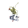
| ||||||||
|---|---|---|---|---|---|---|---|---|---|
| 1 |
| ||||||||
| 2 | 
| ||||||||
| 3 | 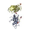
| ||||||||
| Unit cell |
|
-Components
| #1: Protein | Mass: 13715.132 Da / Num. of mol.: 1 / Fragment: SH2 DOMAIN (UNP RESIDUES 112-232) Source method: isolated from a genetically manipulated source Source: (gene. exp.) Homo sapiens (human) / Gene: ABL, ABL1, JTK7 / Plasmid: PHFT2 / Production host:  Escherichia coli (E. coli) / Strain (production host): BL21(DE3) Escherichia coli (E. coli) / Strain (production host): BL21(DE3)References: UniProt: P00519, non-specific protein-tyrosine kinase | ||||
|---|---|---|---|---|---|
| #2: Protein | Mass: 11286.290 Da / Num. of mol.: 1 Source method: isolated from a genetically manipulated source Source: (gene. exp.) Homo sapiens (human) / Plasmid: PHFT2 / Production host: Escherichia coli (E. coli) / Strain (production host): BL21(DE3) | ||||
| #3: Chemical | ChemComp-GOL / Glycerol  Mass: 92.094 Da / Num. of mol.: 4 / Source method: obtained synthetically / Formula: C3H8O3 Mass: 92.094 Da / Num. of mol.: 4 / Source method: obtained synthetically / Formula: C3H8O3#4: Chemical | ChemComp-SO4 / | Sulfate  Mass: 96.063 Da / Num. of mol.: 1 / Source method: obtained synthetically / Formula: SO4 Mass: 96.063 Da / Num. of mol.: 1 / Source method: obtained synthetically / Formula: SO4#5: Water | ChemComp-HOH / | Water Mass: 18.015 Da / Num. of mol.: 127 / Source method: isolated from a natural source / Formula: H2O Mass: 18.015 Da / Num. of mol.: 127 / Source method: isolated from a natural source / Formula: H2O |
-Experimental details
-Experiment
| Experiment | Method: X-RAY DIFFRACTION |
|---|
- Sample preparation
| Crystal | Density Matthews: 2.09 Å3/Da / Density % sol: 41.03 % |
|---|---|
| Crystal grow | pH: 6 Details: 0.2M MG(NO3)2, 100MM LICL, 20% PEG 3350, VAPOR DIFFUSION, HANGING DROP, TEMPERATURE 292K, pH 6.0 |
-Data collection
| Diffraction | Mean temperature: 100 K |
|---|---|
| Diffraction source | Source: SYNCHROTRON / Site: APS  / Beamline: 24-ID-E / Wavelength: 0.97917 / Beamline: 24-ID-E / Wavelength: 0.97917 |
| Detector | Type: ADSC QUANTUM 315 / Detector: CCD / Date: Mar 5, 2010 |
| Radiation | Monochromator: SI 111 SIDE BOUNCE MONOCHROMATOR / Protocol: SINGLE WAVELENGTH / Monochromatic (M) / Laue (L): M / Scattering type: x-ray |
| Radiation wavelength | Wavelength: 0.97917 Å / Relative weight: 1 |
| Reflection | Resolution: 2.1→30 Å / Num. obs: 12700 / % possible obs: 99.2 % / Observed criterion σ(I): 2 / Redundancy: 5.2 % / Rsym value: 0.133 / Net I/σ(I): 11.8 |
| Reflection shell | Resolution: 2.1→2.18 Å / Redundancy: 5.1 % / Mean I/σ(I) obs: 2.4 / % possible all: 99.3 |
- Processing
| Software |
| |||||||||||||||||||||||||||||||||||||||||||||||||||||||||||||||||
|---|---|---|---|---|---|---|---|---|---|---|---|---|---|---|---|---|---|---|---|---|---|---|---|---|---|---|---|---|---|---|---|---|---|---|---|---|---|---|---|---|---|---|---|---|---|---|---|---|---|---|---|---|---|---|---|---|---|---|---|---|---|---|---|---|---|---|
| Refinement | Method to determine structure: MOLECULAR REPLACEMENT Starting model: 1OPK: 154-235, 1FNF: LOOPS OMITTED Resolution: 2.1→30 Å / Cor.coef. Fo:Fc: 0.942 / Cor.coef. Fo:Fc free: 0.902 / SU B: 4.957 / SU ML: 0.133 / Cross valid method: THROUGHOUT / σ(F): 2 / ESU R Free: 0.205 / Stereochemistry target values: MAXIMUM LIKELIHOOD / Details: HYDROGENS HAVE BEEN ADDED IN THE RIDING POSITIONS
| |||||||||||||||||||||||||||||||||||||||||||||||||||||||||||||||||
| Solvent computation | Ion probe radii: 0.8 Å / Shrinkage radii: 0.8 Å / VDW probe radii: 1.4 Å / Solvent model: MASK | |||||||||||||||||||||||||||||||||||||||||||||||||||||||||||||||||
| Displacement parameters | Biso mean: 24.52 Å2
| |||||||||||||||||||||||||||||||||||||||||||||||||||||||||||||||||
| Refinement step | Cycle: LAST / Resolution: 2.1→30 Å
| |||||||||||||||||||||||||||||||||||||||||||||||||||||||||||||||||
| Refine LS restraints |
| |||||||||||||||||||||||||||||||||||||||||||||||||||||||||||||||||
| LS refinement shell | Resolution: 2.1→2.15 Å / Total num. of bins used: 20
|
