 Movie
Movie Controller


+ Open data

- Basic information
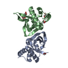
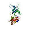
| Entry | Database: PDB / ID: 4gr6 | ||||||
|---|---|---|---|---|---|---|---|
| Title | Crystal structure of AtRbcX2 from Arabidopsis thaliana | ||||||
 Components Components | AtRbcX2 | ||||||
 Keywords Keywords |  CHAPERONE / Rubisco / RbcX CHAPERONE / Rubisco / RbcX | ||||||
| Function / homology |  Function and homology informationchloroplast stroma / chaperone-mediated protein folding / protein folding chaperone Function and homology informationchloroplast stroma / chaperone-mediated protein folding / protein folding chaperoneSimilarity search - Function | ||||||
| Biological species |  Arabidopsis thaliana (thale cress) Arabidopsis thaliana (thale cress) | ||||||
| Method | X-RAY DIFFRACTION / MOLECULAR REPLACEMENT / Resolution: 2 Å | ||||||
 Authors Authors | Grudnik, P. / Golik, P. / Kolesinski, P. / Dubin, G. / Szczepaniak, A. | ||||||
 Citation Citation | Journal: Biochim.Biophys.Acta / Year: 2013 Title: Insights into eukaryotic Rubisco assembly - Crystal structures of RbcX chaperones from Arabidopsis thaliana. Authors: Kolesinski, P. / Golik, P. / Grudnik, P. / Piechota, J. / Markiewicz, M. / Tarnawski, M. / Dubin, G. / Szczepaniak, A. | ||||||
| History |
|
- Structure visualization
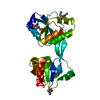
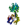
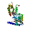
| Structure viewer | Molecule: MolmilJmol/JSmol |
|---|
- Downloads & links
-Download
| PDBx/mmCIF format | 4gr6.cif.gz | 105 KB | Display | PDBx/mmCIF format |
|---|---|---|---|---|
| PDB format | pdb4gr6.ent.gz | 81.2 KB | Display | PDB format |
| PDBx/mmJSON format | 4gr6.json.gz | Tree view | PDBx/mmJSON format | |
| Others |  Other downloads Other downloads |
-Validation report
| Arichive directory | https://data.pdbj.org/pub/pdb/validation_reports/gr/4gr6ftp://data.pdbj.org/pub/pdb/validation_reports/gr/4gr6 | HTTPS FTP |
|---|
-Related structure data
| Related structure data | 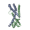 4gr2C 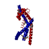 2z44S C: citing same article ( S: Starting model for refinement |
|---|---|
| Similar structure data |


-Links
 PDBj
PDBj

- Assembly
| Deposited unit | 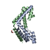
| ||||||||
|---|---|---|---|---|---|---|---|---|---|
| 1 |
| ||||||||
| Unit cell |
| ||||||||
| Components on special symmetry positions |
|
-Components
| #1: Protein | Mass: 15039.025 Da / Num. of mol.: 2 / Fragment: UNP residues 79-203 / Mutation: C91A Source method: isolated from a genetically manipulated source Source: (gene. exp.) Arabidopsis thaliana (thale cress) / Gene: At5g19855 / Production host:  Escherichia coli (E. coli) / References: UniProt: Q8L9X2 Escherichia coli (E. coli) / References: UniProt: Q8L9X2#2: Chemical | ChemComp-EDO / | Ethylene glycol  Mass: 62.068 Da / Num. of mol.: 1 / Source method: obtained synthetically / Formula: C2H6O2 Mass: 62.068 Da / Num. of mol.: 1 / Source method: obtained synthetically / Formula: C2H6O2#3: Water | ChemComp-HOH / | Water Mass: 18.015 Da / Num. of mol.: 91 / Source method: isolated from a natural source / Formula: H2O Mass: 18.015 Da / Num. of mol.: 91 / Source method: isolated from a natural source / Formula: H2O |
|---|
-Experimental details
-Experiment
| Experiment | Method: X-RAY DIFFRACTION / Number of used crystals: 1 |
|---|
- Sample preparation
| Crystal | Density Matthews: 2.75 Å3/Da / Density % sol: 55.26 % |
|---|---|
| Crystal grow | Temperature: 292 K / Method: vapor diffusion, sitting drop / pH: 5.6 Details: 15% PEG 400, 100 mM sodium citrate , pH 5.6, VAPOR DIFFUSION, SITTING DROP, temperature 292K |
-Data collection
| Diffraction | Mean temperature: 100 K |
|---|---|
| Diffraction source | Source: ROTATING ANODE / Type: RIGAKU MICROMAX-007 HF / Wavelength: 1.54 Å |
| Detector | Type: RIGAKU RAXIS IV++ / Detector: IMAGE PLATE / Date: May 21, 2012 |
| Radiation | Protocol: SINGLE WAVELENGTH / Monochromatic (M) / Laue (L): M / Scattering type: x-ray |
| Radiation wavelength | Wavelength: 1.54 Å / Relative weight: 1 |
| Reflection | Resolution: 2→33.75 Å / Num. all: 21808 / Num. obs: 21808 / % possible obs: 100 % |
- Processing
| Software |
| |||||||||||||||||||||||||||||||||||||||||||||||||||||||||||||||
|---|---|---|---|---|---|---|---|---|---|---|---|---|---|---|---|---|---|---|---|---|---|---|---|---|---|---|---|---|---|---|---|---|---|---|---|---|---|---|---|---|---|---|---|---|---|---|---|---|---|---|---|---|---|---|---|---|---|---|---|---|---|---|---|---|
| Refinement | Method to determine structure: MOLECULAR REPLACEMENT Starting model: 2Z44 Resolution: 2→21.167 Å / SU ML: 0.16 / σ(F): 1.35 / Phase error: 21.03 / Stereochemistry target values: ML
| |||||||||||||||||||||||||||||||||||||||||||||||||||||||||||||||
| Solvent computation | Shrinkage radii: 0.9 Å / VDW probe radii: 1.11 Å / Solvent model: FLAT BULK SOLVENT MODEL | |||||||||||||||||||||||||||||||||||||||||||||||||||||||||||||||
| Refinement step | Cycle: LAST / Resolution: 2→21.167 Å
| |||||||||||||||||||||||||||||||||||||||||||||||||||||||||||||||
| Refine LS restraints |
| |||||||||||||||||||||||||||||||||||||||||||||||||||||||||||||||
| LS refinement shell |
| |||||||||||||||||||||||||||||||||||||||||||||||||||||||||||||||
| Refinement TLS params. | Method: refined / Origin x: -12.1395 Å / Origin y: 19.2645 Å / Origin z: -14.6886 Å
| |||||||||||||||||||||||||||||||||||||||||||||||||||||||||||||||
| Refinement TLS group | Selection details: all |
