 Movie
Movie Controller


+ Open data

- Basic information
| Entry | Database: PDB / ID: 2rbi | ||||||
|---|---|---|---|---|---|---|---|
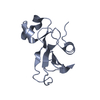
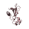
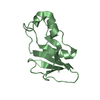
| Title | STRUCTURE OF BINASE MUTANT HIS 101 ASN | ||||||
 Components Components | RIBONUCLEASE | ||||||
 Keywords Keywords | ENDORIBONUCLEASE / MICROBIAL EXTRACELLULAR RIBONUCLEASE / PHOSPHODIESTER TRANSFERASE / HYDROLASE | ||||||
| Function / homology |  Function and homology informationHydrolases; Acting on ester bonds; Endoribonucleases producing 3'-phosphomonoesters / RNA endonuclease activity / RNA binding / extracellular region Function and homology informationHydrolases; Acting on ester bonds; Endoribonucleases producing 3'-phosphomonoesters / RNA endonuclease activity / RNA binding / extracellular regionSimilarity search - Function | ||||||
| Biological species |  | ||||||
| Method | X-RAY DIFFRACTION / MOLECULAR REPLACEMENT / Resolution: 2.2 Å | ||||||
 Authors Authors | Offen, W.A. / Okorokov, A.L. | ||||||
 Citation Citation | Journal: Protein Eng. / Year: 1997 Title: RNA cleavage without hydrolysis. Splitting the catalytic activities of binase with Asn101 and Thr101 mutations. Authors: Okorokov, A.L. / Panov, K.I. / Offen, W.A. / Mukhortov, V.G. / Antson, A.A. / Karpeisky, M.Y.a. / Wilkinson, A.J. / Dodson, G.G. | ||||||
| History |
|
- Structure visualization
| Structure viewer | Molecule: MolmilJmol/JSmol |
|---|
- Downloads & links
-Download
| PDBx/mmCIF format | 2rbi.cif.gz | 59.8 KB | Display | PDBx/mmCIF format |
|---|---|---|---|---|
| PDB format | pdb2rbi.ent.gz | 44.1 KB | Display | PDB format |
| PDBx/mmJSON format | 2rbi.json.gz | Tree view | PDBx/mmJSON format | |
| Others |  Other downloads Other downloads |
-Validation report
| Arichive directory | https://data.pdbj.org/pub/pdb/validation_reports/rb/2rbiftp://data.pdbj.org/pub/pdb/validation_reports/rb/2rbi | HTTPS FTP |
|---|
-Related structure data
| Similar structure data |
|---|


-Links
 PDBj
PDBj- Assembly
| Deposited unit | 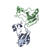
| ||||||||
|---|---|---|---|---|---|---|---|---|---|
| 1 | 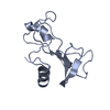
| ||||||||
| 2 | 
| ||||||||
| Unit cell |
|
-Components
| #1: Protein | / BINASE / EXTRACELLULAR RIBONUCLEASE FROM BACILLUS INTERMEDIUS Mass: 12203.579 Da / Num. of mol.: 2 / Mutation: H101N Source method: isolated from a genetically manipulated source Source: (gene. exp.) Description: BINASE GENE CO-EXPRESSED AS FUSION PROTEIN WITH ALKALINE PHOSPHATASE TO ENABLE EXPORT INTO PERIPLASMIC SPACE Gene: PHOA-BINASE HIS101ASN / Plasmid: PEXBINN101 / Gene (production host): PHOA-BINASE HIS101ASN / Production host: Escherichia coli (E. coli)Strain (production host): EPICURIAN COLI STRAIN XL-1 BLUE MRF' References: UniProt: P00649, Hydrolases; Acting on ester bonds; Endoribonucleases producing 3'-phosphomonoesters#2: Water | ChemComp-HOH / | Water Mass: 18.015 Da / Num. of mol.: 229 / Source method: isolated from a natural source / Formula: H2O Mass: 18.015 Da / Num. of mol.: 229 / Source method: isolated from a natural source / Formula: H2O |
|---|
-Experimental details
-Experiment
| Experiment | Method: X-RAY DIFFRACTION / Number of used crystals: 1 |
|---|
- Sample preparation
| Crystal | Density Matthews: 2.6 Å3/Da / Density % sol: 52.6 % | ||||||||||||||||||||||||||||||||||||||||
|---|---|---|---|---|---|---|---|---|---|---|---|---|---|---|---|---|---|---|---|---|---|---|---|---|---|---|---|---|---|---|---|---|---|---|---|---|---|---|---|---|---|
| Crystal grow | Temperature: 291 K / Method: vapor diffusion / pH: 7.5 Details: VAPOR DIFFUSION METHOD, WITH 12MG/ML BINASE HIS101ASN, 40MM GLYCINE PH 7.5, 8.75% POLYETHYL- ENE GLYCOL MR 10,000 AND 2.5% SATURATED SODIUM CITRATE IN THE HANGING DROP, AND 17.5% PEG 10,000, ...Details: VAPOR DIFFUSION METHOD, WITH 12MG/ML BINASE HIS101ASN, 40MM GLYCINE PH 7.5, 8.75% POLYETHYL- ENE GLYCOL MR 10,000 AND 2.5% SATURATED SODIUM CITRATE IN THE HANGING DROP, AND 17.5% PEG 10,000, 60MM GLYCINE PH 7.5 AND 5% SODIUM CITRATE IN WELL. LEFT FOR 2 DAYS AT 18 DEGREES CENTIGRADE., vapor diffusion, temperature 291K | ||||||||||||||||||||||||||||||||||||||||
| Crystal grow | *PLUS Method: vapor diffusion, hanging drop | ||||||||||||||||||||||||||||||||||||||||
| Components of the solutions | *PLUS
|
-Data collection
| Diffraction | Mean temperature: 291 K |
|---|---|
| Diffraction source | Source: ROTATING ANODE / Type: RIGAKU RUH2R / Wavelength: 1.5418 |
| Detector | Type: RIGAKU / Detector: IMAGE PLATE / Date: Oct 22, 1993 |
| Radiation | Monochromator: GRAPHITE(002) / Monochromatic (M) / Laue (L): M / Scattering type: x-ray |
| Radiation wavelength | Wavelength: 1.5418 Å / Relative weight: 1 |
| Reflection | Resolution: 2.2→20 Å / Num. obs: 13288 / % possible obs: 97.2 % / Observed criterion σ(I): 0 / Redundancy: 3.9 % / Biso Wilson estimate: 19.6 Å2 / Rmerge(I) obs: 0.055 / Net I/σ(I): 7.5 |
| Reflection shell | Resolution: 2.2→2.26 Å / Redundancy: 2.9 % / Rmerge(I) obs: 0.196 / Mean I/σ(I) obs: 3.8 / % possible all: 80.7 |
| Reflection | *PLUS Num. measured all: 51822 |
| Reflection shell | *PLUS % possible obs: 80.7 % |
- Processing
| Software |
| ||||||||||||||||||||||||||||||||||||||||||||||||||||||||||||||||||||||||||||||||||||
|---|---|---|---|---|---|---|---|---|---|---|---|---|---|---|---|---|---|---|---|---|---|---|---|---|---|---|---|---|---|---|---|---|---|---|---|---|---|---|---|---|---|---|---|---|---|---|---|---|---|---|---|---|---|---|---|---|---|---|---|---|---|---|---|---|---|---|---|---|---|---|---|---|---|---|---|---|---|---|---|---|---|---|---|---|---|
| Refinement | Method to determine structure: MOLECULAR REPLACEMENT Starting model: WILD-TYPE BINASE Resolution: 2.2→10 Å / σ(F): 0
| ||||||||||||||||||||||||||||||||||||||||||||||||||||||||||||||||||||||||||||||||||||
| Displacement parameters | Biso mean: 27.4 Å2 | ||||||||||||||||||||||||||||||||||||||||||||||||||||||||||||||||||||||||||||||||||||
| Refinement step | Cycle: LAST / Resolution: 2.2→10 Å
| ||||||||||||||||||||||||||||||||||||||||||||||||||||||||||||||||||||||||||||||||||||
| Refine LS restraints |
| ||||||||||||||||||||||||||||||||||||||||||||||||||||||||||||||||||||||||||||||||||||
| Software | *PLUS Name: PROLSQ / Classification: refinement | ||||||||||||||||||||||||||||||||||||||||||||||||||||||||||||||||||||||||||||||||||||
| Refinement | *PLUS Rfactor obs: 0.178 | ||||||||||||||||||||||||||||||||||||||||||||||||||||||||||||||||||||||||||||||||||||
| Solvent computation | *PLUS | ||||||||||||||||||||||||||||||||||||||||||||||||||||||||||||||||||||||||||||||||||||
| Displacement parameters | *PLUS |
