 Movie
Movie Controller


+ Open data

- Basic information
| Entry | Database: PDB / ID: 4goy | ||||||
|---|---|---|---|---|---|---|---|
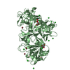
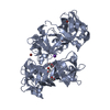
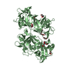
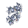
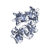
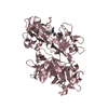
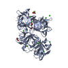
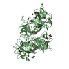
| Title | The crystal structure of human fascin 1 K41A mutant | ||||||
 Components Components | Fascin | ||||||
 Keywords Keywords | PROTEIN BINDING / beta-trefoil / actin bundling protein / cancer / metastasis / cell migration / Actin-binding / Phosphoprotein / Actin | ||||||
| Function / homology |  Function and homology information Function and homology informationmicrospike / parallel actin filament bundle assembly / regulation of microvillus assembly / positive regulation of extracellular matrix disassembly / establishment of apical/basal cell polarity / microspike assembly / cell projection membrane / podosome / positive regulation of podosome assembly / cell-cell junction assembly ...microspike / parallel actin filament bundle assembly / regulation of microvillus assembly / positive regulation of extracellular matrix disassembly / establishment of apical/basal cell polarity / microspike assembly / cell projection membrane / podosome / positive regulation of podosome assembly / cell-cell junction assembly / positive regulation of filopodium assembly / establishment or maintenance of cell polarity / microvillus / actin filament bundle assembly / positive regulation of lamellipodium assembly / stress fiber / ruffle / filopodium / cell motility / regulation of actin cytoskeleton organization / actin filament binding / cell-cell junction / cell migration / actin cytoskeleton / lamellipodium / protein-macromolecule adaptor activity / actin binding / cell cortex / growth cone / actin cytoskeleton organization / Interleukin-4 and Interleukin-13 signaling / cytoskeleton / cadherin binding / RNA binding / extracellular exosome / cytosol / cytoplasmSimilarity search - Function | ||||||
| Biological species |  Homo sapiens (human) Homo sapiens (human) | ||||||
| Method | X-RAY DIFFRACTION / SYNCHROTRON / MOLECULAR REPLACEMENT / Resolution: 2.3 Å | ||||||
 Authors Authors | Yang, S.Y. / Huang, F.K. / Huang, J. / Chen, S. / Jakoncic, J. / Leo-Macias, A. / Diaz-Avalos, R. / Chen, L. / Zhang, J.J. / Huang, X.Y. | ||||||
 Citation Citation | Journal: J.Biol.Chem. / Year: 2013 Title: Molecular mechanism of fascin function in filopodial formation. Authors: Yang, S. / Huang, F.K. / Huang, J. / Chen, S. / Jakoncic, J. / Leo-Macias, A. / Diaz-Avalos, R. / Chen, L. / Zhang, J.J. / Huang, X.Y. | ||||||
| History |
|
- Structure visualization
| Structure viewer | Molecule: MolmilJmol/JSmol |
|---|
- Downloads & links
-Download
| PDBx/mmCIF format | 4goy.cif.gz | 393.1 KB | Display | PDBx/mmCIF format |
|---|---|---|---|---|
| PDB format | pdb4goy.ent.gz | 323.5 KB | Display | PDB format |
| PDBx/mmJSON format | 4goy.json.gz | Tree view | PDBx/mmJSON format | |
| Others |  Other downloads Other downloads |
-Validation report
| Arichive directory | https://data.pdbj.org/pub/pdb/validation_reports/go/4goyftp://data.pdbj.org/pub/pdb/validation_reports/go/4goy | HTTPS FTP |
|---|
-Related structure data
| Related structure data | 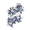 4govC  4gp0C  4gp3C  3llpS C: citing same article ( S: Starting model for refinement |
|---|---|
| Similar structure data |


-Links
 PDBj
PDBj


- Assembly
| Deposited unit | 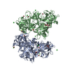
| ||||||||
|---|---|---|---|---|---|---|---|---|---|
| 1 | 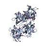
| ||||||||
| 2 | 
| ||||||||
| 3 |
| ||||||||
| 4 | 
| ||||||||
| Unit cell |
|
-Components
-Protein , 1 types, 2 molecules AB
| #1: Protein | / 55 kDa actin-bundling protein / Singed-like protein / p55 Mass: 54543.773 Da / Num. of mol.: 2 / Mutation: K41A Source method: isolated from a genetically manipulated source Source: (gene. exp.) Homo sapiens (human) / Gene: FAN1, FSCN1, HSN, SNL / Plasmid: pGEX4T-Fascin1 / Production host:  Escherichia coli (E. coli) / Strain (production host): BL21(DE3) / References: UniProt: Q16658 Escherichia coli (E. coli) / Strain (production host): BL21(DE3) / References: UniProt: Q16658 |
|---|
-Non-polymers , 5 types, 476 molecules 


| #2: Chemical | ChemComp-BR / Bromide Mass: 79.904 Da / Num. of mol.: 8 / Source method: obtained synthetically / Formula: Br Mass: 79.904 Da / Num. of mol.: 8 / Source method: obtained synthetically / Formula: Br#3: Chemical | ChemComp-CL / Chloride Mass: 35.453 Da / Num. of mol.: 10 / Source method: obtained synthetically / Formula: Cl Mass: 35.453 Da / Num. of mol.: 10 / Source method: obtained synthetically / Formula: Cl#4: Chemical | ChemComp-GOL / Glycerol Mass: 92.094 Da / Num. of mol.: 6 / Source method: obtained synthetically / Formula: C3H8O3 Mass: 92.094 Da / Num. of mol.: 6 / Source method: obtained synthetically / Formula: C3H8O3#5: Chemical | ChemComp-DTT / | Dithiothreitol Mass: 154.251 Da / Num. of mol.: 1 / Source method: obtained synthetically / Formula: C4H10O2S2 Mass: 154.251 Da / Num. of mol.: 1 / Source method: obtained synthetically / Formula: C4H10O2S2#6: Water | ChemComp-HOH / | WaterMass: 18.015 Da / Num. of mol.: 451 / Source method: isolated from a natural source / Formula: H2O |
|---|
-Experimental details
-Experiment
| Experiment | Method: X-RAY DIFFRACTION / Number of used crystals: 1 |
|---|
- Sample preparation
| Crystal | Density Matthews: 2.2 Å3/Da / Density % sol: 43.98 % |
|---|---|
| Crystal grow | Temperature: 293 K / Method: vapor diffusion, hanging drop / pH: 8 Details: 100mM Hepes, 16% PEG 4000, 1% isopropanol , pH 8.0, VAPOR DIFFUSION, HANGING DROP, temperature 293K |
-Data collection
| Diffraction | Mean temperature: 100 K |
|---|---|
| Diffraction source | Source: SYNCHROTRON / Site: NSLS  / Beamline: X6A / Wavelength: 1 Å / Beamline: X6A / Wavelength: 1 Å |
| Detector | Type: ADSC QUANTUM 210 / Detector: CCD / Date: Oct 25, 2010 |
| Radiation | Monochromator: Si 111 CHANNEL CUT / Protocol: SINGLE WAVELENGTH / Monochromatic (M) / Laue (L): M / Scattering type: x-ray |
| Radiation wavelength | Wavelength: 1 Å / Relative weight: 1 |
| Reflection | Resolution: 2.3→30 Å / Num. obs: 42281 / % possible obs: 99.8 % / Observed criterion σ(I): 2 / Redundancy: 3.6 % / Rmerge(I) obs: 0.065 / Net I/σ(I): 24.71 |
| Reflection shell | Resolution: 2.3→2.34 Å / Redundancy: 3.6 % / Rmerge(I) obs: 0.39 / Mean I/σ(I) obs: 4.51 / Num. unique all: 2086 / % possible all: 100 |
- Processing
| Software |
| ||||||||||||||||||||||||||||||||||||||||||||||||||||||||||||||||||||||||||||||||||||||||||||||||||||||||||||||||||||||||||||||||||||||||||||||||||||||||||||||||||||||||||||||||||||||||||||||||||||||||||||||||||||||||||||||||||||||||||||||||||||||||||
|---|---|---|---|---|---|---|---|---|---|---|---|---|---|---|---|---|---|---|---|---|---|---|---|---|---|---|---|---|---|---|---|---|---|---|---|---|---|---|---|---|---|---|---|---|---|---|---|---|---|---|---|---|---|---|---|---|---|---|---|---|---|---|---|---|---|---|---|---|---|---|---|---|---|---|---|---|---|---|---|---|---|---|---|---|---|---|---|---|---|---|---|---|---|---|---|---|---|---|---|---|---|---|---|---|---|---|---|---|---|---|---|---|---|---|---|---|---|---|---|---|---|---|---|---|---|---|---|---|---|---|---|---|---|---|---|---|---|---|---|---|---|---|---|---|---|---|---|---|---|---|---|---|---|---|---|---|---|---|---|---|---|---|---|---|---|---|---|---|---|---|---|---|---|---|---|---|---|---|---|---|---|---|---|---|---|---|---|---|---|---|---|---|---|---|---|---|---|---|---|---|---|---|---|---|---|---|---|---|---|---|---|---|---|---|---|---|---|---|---|---|---|---|---|---|---|---|---|---|---|---|---|---|---|---|---|---|---|---|---|---|---|---|---|---|---|---|---|---|---|---|---|
| Refinement | Method to determine structure: MOLECULAR REPLACEMENT Starting model: 3LLP Resolution: 2.3→30 Å / Cor.coef. Fo:Fc: 0.944 / Cor.coef. Fo:Fc free: 0.919 / SU B: 16.024 / SU ML: 0.192 / Cross valid method: THROUGHOUT / ESU R: 0.467 / ESU R Free: 0.256 / Stereochemistry target values: MAXIMUM LIKELIHOOD / Details: HYDROGENS HAVE BEEN USED IF PRESENT IN THE INPUT
| ||||||||||||||||||||||||||||||||||||||||||||||||||||||||||||||||||||||||||||||||||||||||||||||||||||||||||||||||||||||||||||||||||||||||||||||||||||||||||||||||||||||||||||||||||||||||||||||||||||||||||||||||||||||||||||||||||||||||||||||||||||||||||
| Solvent computation | Ion probe radii: 0.8 Å / Shrinkage radii: 0.8 Å / VDW probe radii: 1.2 Å / Solvent model: MASK | ||||||||||||||||||||||||||||||||||||||||||||||||||||||||||||||||||||||||||||||||||||||||||||||||||||||||||||||||||||||||||||||||||||||||||||||||||||||||||||||||||||||||||||||||||||||||||||||||||||||||||||||||||||||||||||||||||||||||||||||||||||||||||
| Displacement parameters | Biso mean: 38.797 Å2
| ||||||||||||||||||||||||||||||||||||||||||||||||||||||||||||||||||||||||||||||||||||||||||||||||||||||||||||||||||||||||||||||||||||||||||||||||||||||||||||||||||||||||||||||||||||||||||||||||||||||||||||||||||||||||||||||||||||||||||||||||||||||||||
| Refinement step | Cycle: LAST / Resolution: 2.3→30 Å
| ||||||||||||||||||||||||||||||||||||||||||||||||||||||||||||||||||||||||||||||||||||||||||||||||||||||||||||||||||||||||||||||||||||||||||||||||||||||||||||||||||||||||||||||||||||||||||||||||||||||||||||||||||||||||||||||||||||||||||||||||||||||||||
| Refine LS restraints |
| ||||||||||||||||||||||||||||||||||||||||||||||||||||||||||||||||||||||||||||||||||||||||||||||||||||||||||||||||||||||||||||||||||||||||||||||||||||||||||||||||||||||||||||||||||||||||||||||||||||||||||||||||||||||||||||||||||||||||||||||||||||||||||
| LS refinement shell | Resolution: 2.291→2.35 Å / Total num. of bins used: 20
| ||||||||||||||||||||||||||||||||||||||||||||||||||||||||||||||||||||||||||||||||||||||||||||||||||||||||||||||||||||||||||||||||||||||||||||||||||||||||||||||||||||||||||||||||||||||||||||||||||||||||||||||||||||||||||||||||||||||||||||||||||||||||||
| Refinement TLS params. | Method: refined / Refine-ID: X-RAY DIFFRACTION
| ||||||||||||||||||||||||||||||||||||||||||||||||||||||||||||||||||||||||||||||||||||||||||||||||||||||||||||||||||||||||||||||||||||||||||||||||||||||||||||||||||||||||||||||||||||||||||||||||||||||||||||||||||||||||||||||||||||||||||||||||||||||||||
| Refinement TLS group |
|
