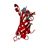
 Movie
Movie Controller


+ Open data

- Basic information
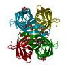
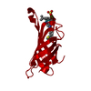
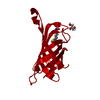
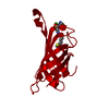
| Entry | Database: PDB / ID: 4gjv | ||||||
|---|---|---|---|---|---|---|---|
| Title | Streptavidin-S112H | ||||||
 Components Components | Streptavidin | ||||||
 Keywords Keywords | Biotin-binding protein / artificial metalloenyzme / artificial transfer hydrogenase / beta barrel / tetramer / biotin / iridium pentamethylcyclopentadienyl | ||||||
| Function / homology |  Function and homology information Function and homology information | ||||||
| Biological species |  Streptomyces avidinii (bacteria) Streptomyces avidinii (bacteria) | ||||||
| Method | X-RAY DIFFRACTION / SYNCHROTRON / MOLECULAR REPLACEMENT / Resolution: 2.4 Å | ||||||
 Authors Authors | Heinisch, T. / Schirmer, T. | ||||||
 Citation Citation | Journal: J.Am.Chem.Soc. / Year: 2013 Title: A dual anchoring strategy for the localization and activation of artificial metalloenzymes based on the biotin-streptavidin technology. Authors: Zimbron, J.M. / Heinisch, T. / Schmid, M. / Hamels, D. / Nogueira, E.S. / Schirmer, T. / Ward, T.R. | ||||||
| History |
|
- Structure visualization
| Structure viewer | Molecule: MolmilJmol/JSmol |
|---|
- Downloads & links
-Download
| PDBx/mmCIF format | 4gjv.cif.gz | 63.3 KB | Display | PDBx/mmCIF format |
|---|---|---|---|---|
| PDB format | pdb4gjv.ent.gz | 45.7 KB | Display | PDB format |
| PDBx/mmJSON format | 4gjv.json.gz | Tree view | PDBx/mmJSON format | |
| Others |  Other downloads Other downloads |
-Validation report
| Arichive directory | https://data.pdbj.org/pub/pdb/validation_reports/gj/4gjvftp://data.pdbj.org/pub/pdb/validation_reports/gj/4gjv | HTTPS FTP |
|---|
-Related structure data
| Related structure data |  4gjsC  2qcbS S: Starting model for refinement C: citing same article ( |
|---|---|
| Similar structure data |


-Links
 PDBj
PDBj- Assembly
| Deposited unit | 
| |||||||||
|---|---|---|---|---|---|---|---|---|---|---|
| 1 | 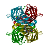
| |||||||||
| Unit cell |
| |||||||||
| Components on special symmetry positions |
|
-Components
| #1: Protein | Mass: 16621.086 Da / Num. of mol.: 1 / Mutation: S112H Source method: isolated from a genetically manipulated source Source: (gene. exp.) Streptomyces avidinii (bacteria) / Plasmid: pLysS / Production host: Escherichia coli (E. coli) / Strain (production host): BL21(DE3) / References: UniProt: P22629 | ||||
|---|---|---|---|---|---|
| #2: Chemical | ChemComp-0OD /   Mass: 599.827 Da / Num. of mol.: 1 / Source method: obtained synthetically / Formula: C21H32Cl3N3O2RhS Mass: 599.827 Da / Num. of mol.: 1 / Source method: obtained synthetically / Formula: C21H32Cl3N3O2RhS | ||||
| #3: Chemical | ChemComp-CL / Chloride  Mass: 35.453 Da / Num. of mol.: 4 / Source method: obtained synthetically / Formula: Cl Mass: 35.453 Da / Num. of mol.: 4 / Source method: obtained synthetically / Formula: Cl#4: Chemical | ChemComp-RH / | Rhodium  Mass: 102.906 Da / Num. of mol.: 1 / Source method: obtained synthetically / Formula: Rh Mass: 102.906 Da / Num. of mol.: 1 / Source method: obtained synthetically / Formula: Rh#5: Water | ChemComp-HOH / | Water Mass: 18.015 Da / Num. of mol.: 22 / Source method: isolated from a natural source / Formula: H2O Mass: 18.015 Da / Num. of mol.: 22 / Source method: isolated from a natural source / Formula: H2O |
-Experimental details
-Experiment
| Experiment | Method: X-RAY DIFFRACTION / Number of used crystals: 1 |
|---|
- Sample preparation
| Crystal | Density Matthews: 2.3 Å3/Da / Density % sol: 46.9 % |
|---|---|
| Crystal grow | Temperature: 277 K / Method: vapor diffusion, hanging drop / pH: 6 Details: 0.1 M MES, 19 % PEG500, pH 6.0, VAPOR DIFFUSION, HANGING DROP, temperature 277K |
-Data collection
| Diffraction | Mean temperature: 100 K |
|---|---|
| Diffraction source | Source: SYNCHROTRON / Site: SLS  / Beamline: X06DA / Wavelength: 1 Å / Beamline: X06DA / Wavelength: 1 Å |
| Detector | Type: MARMOSAIC 225 mm CCD / Detector: CCD / Date: Jun 3, 2011 |
| Radiation | Protocol: SINGLE WAVELENGTH / Monochromatic (M) / Laue (L): M / Scattering type: x-ray |
| Radiation wavelength | Wavelength: 1 Å / Relative weight: 1 |
| Reflection | Resolution: 2.4→54.946 Å / Num. all: 11694 / Num. obs: 10084 |
- Processing
| Software |
| ||||||||||||||||||||||||||||||||||||||||
|---|---|---|---|---|---|---|---|---|---|---|---|---|---|---|---|---|---|---|---|---|---|---|---|---|---|---|---|---|---|---|---|---|---|---|---|---|---|---|---|---|---|
| Refinement | Method to determine structure: MOLECULAR REPLACEMENT Starting model: PDB ENTRY 2QCB Resolution: 2.4→19.876 Å / Occupancy max: 1 / Occupancy min: 0 / SU ML: 0.32 / σ(F): 1.34 / Phase error: 25.05 / Stereochemistry target values: ML
| ||||||||||||||||||||||||||||||||||||||||
| Solvent computation | Shrinkage radii: 0.9 Å / VDW probe radii: 1.11 Å / Solvent model: FLAT BULK SOLVENT MODEL | ||||||||||||||||||||||||||||||||||||||||
| Displacement parameters | Biso max: 83.66 Å2 / Biso mean: 35.641 Å2 / Biso min: 20.01 Å2 | ||||||||||||||||||||||||||||||||||||||||
| Refinement step | Cycle: LAST / Resolution: 2.4→19.876 Å
| ||||||||||||||||||||||||||||||||||||||||
| Refinement TLS params. | Method: refined / Origin x: 14.4174 Å / Origin y: 24.8201 Å / Origin z: -3.3717 Å
| ||||||||||||||||||||||||||||||||||||||||
| Refinement TLS group | Selection details: chain A and resid 13:134 |
