 Movie
Movie Controller

[English] 日本語
 Yorodumi
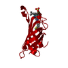
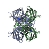
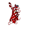
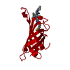
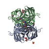
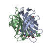
- PDB-2qcb: T7-tagged full-length streptavidin complexed with ruthenium ligand -
+ Open data

- Basic information
| Entry | Database: PDB / ID: 2qcb | ||||||
|---|---|---|---|---|---|---|---|
| Title | T7-tagged full-length streptavidin complexed with ruthenium ligand | ||||||
 Components Components | Streptavidin | ||||||
 Keywords Keywords | BIOTIN BINDING PROTEIN / streptavidin / t7-tag / artificial transfer hydrogenase | ||||||
| Function / homology |  Function and homology information Function and homology information | ||||||
| Biological species |  Streptomyces avidinii (bacteria) Streptomyces avidinii (bacteria) | ||||||
| Method | X-RAY DIFFRACTION / MOLECULAR REPLACEMENT / Resolution: 1.65 Å | ||||||
 Authors Authors | Le Trong, I. / Creus, M. / Pordea, A. / Ward, T.R. / Stenkamp, R.E. | ||||||
 Citation Citation | Journal: Angew.Chem.Int.Ed.Engl. / Year: 2008 Title: X-ray structure and designed evolution of an artificial transfer hydrogenase Authors: Creus, M. / Pordea, A. / Rossel, T. / Sardo, A. / Letondor, C. / Ivanova, A. / Letrong, I. / Stenkamp, R.E. / Ward, T.R. | ||||||
| History |
| ||||||
| Remark 600 | HETEROGEN The ligand KYS has considerable molecular motion and disorder. The Ruthenium metal is not ...HETEROGEN The ligand KYS has considerable molecular motion and disorder. The Ruthenium metal is not modeled in confomer B. The SO2N group attached to the linker has rotational disorder and has been modeled as conformer C. |
- Structure visualization
| Structure viewer | Molecule: MolmilJmol/JSmol |
|---|
- Downloads & links
-Download
| PDBx/mmCIF format | 2qcb.cif.gz | 66.6 KB | Display | PDBx/mmCIF format |
|---|---|---|---|---|
| PDB format | pdb2qcb.ent.gz | 49.1 KB | Display | PDB format |
| PDBx/mmJSON format | 2qcb.json.gz | Tree view | PDBx/mmJSON format | |
| Others |  Other downloads Other downloads |
-Validation report
| Arichive directory | https://data.pdbj.org/pub/pdb/validation_reports/qc/2qcbftp://data.pdbj.org/pub/pdb/validation_reports/qc/2qcb | HTTPS FTP |
|---|
-Related structure data
| Related structure data | |
|---|---|
| Similar structure data |


-Links
 PDBj
PDBj- Assembly
| Deposited unit | 
| ||||||||||||
|---|---|---|---|---|---|---|---|---|---|---|---|---|---|
| 1 | 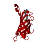
| ||||||||||||
| Unit cell |
| ||||||||||||
| Components on special symmetry positions |
| ||||||||||||
| Details | The biological assembly is a tetramer generated by the deposited chain and three additional subunits generated by application of these crystallographic symmetry operations: 1-x,1-y,z; y,x,-z; 1-y,1-x,-z. |
-Components
| #1: Protein | Mass: 16612.119 Da / Num. of mol.: 1 / Mutation: S112K Source method: isolated from a genetically manipulated source Source: (gene. exp.) Streptomyces avidinii (bacteria) / Plasmid: Pet11B / Production host: Escherichia coli (E. coli) / References: UniProt: P22629 |
|---|---|
| #2: Chemical | ChemComp-KYS /   Mass: 654.187 Da / Num. of mol.: 1 / Source method: obtained synthetically / Formula: C24H31ClN5O4RuS2 Mass: 654.187 Da / Num. of mol.: 1 / Source method: obtained synthetically / Formula: C24H31ClN5O4RuS2 |
| #3: Water | ChemComp-HOH / Water Mass: 18.015 Da / Num. of mol.: 112 / Source method: isolated from a natural source / Formula: H2O Mass: 18.015 Da / Num. of mol.: 112 / Source method: isolated from a natural source / Formula: H2O |
-Experimental details
-Experiment
| Experiment | Method: X-RAY DIFFRACTION / Number of used crystals: 1 |
|---|
- Sample preparation
| Crystal | Density Matthews: 2.3 Å3/Da / Density % sol: 46.48 % |
|---|---|
| Crystal grow | Temperature: 298 K / Method: vapor diffusion, hanging drop / pH: 6.5 Details: protein, 26 mg/ml in water. 4-5x molar excess of ligand. reservoir, 1.0 M sodium citrate, 0.1 M cacodylate buffer, pH 6.5 , VAPOR DIFFUSION, HANGING DROP, temperature 298K |
-Data collection
| Diffraction | Mean temperature: 100 K |
|---|---|
| Diffraction source | Source: ROTATING ANODE / Type: RIGAKU MICROMAX-007 / Wavelength: 1.5418 Å |
| Detector | Type: RIGAKU RAXIS IV / Detector: IMAGE PLATE / Date: May 15, 2006 / Details: mirrors |
| Radiation | Monochromator: mirrors / Protocol: SINGLE WAVELENGTH / Monochromatic (M) / Laue (L): M / Scattering type: x-ray |
| Radiation wavelength | Wavelength: 1.5418 Å / Relative weight: 1 |
| Reflection | Resolution: 1.53→50 Å / Num. all: 21478 / Num. obs: 21478 / % possible obs: 89.3 % / Observed criterion σ(F): 0 / Redundancy: 10.7 % / Biso Wilson estimate: 23 Å2 / Rmerge(I) obs: 0.034 / Χ2: 1.069 / Net I/σ(I): 63.4 |
| Reflection shell | Resolution: 1.53→1.58 Å / Redundancy: 2.2 % / Rmerge(I) obs: 0.256 / Mean I/σ(I) obs: 3.6 / Num. unique all: 650 / Χ2: 1.069 / % possible all: 27.8 |
- Processing
| Software |
| ||||||||||||||||||||||||||||||||||||||||||||||||||||||||||||||||||||||||||||||||||||||||||||||||||||||||||||||||||||||||||||||||||||||||||||
|---|---|---|---|---|---|---|---|---|---|---|---|---|---|---|---|---|---|---|---|---|---|---|---|---|---|---|---|---|---|---|---|---|---|---|---|---|---|---|---|---|---|---|---|---|---|---|---|---|---|---|---|---|---|---|---|---|---|---|---|---|---|---|---|---|---|---|---|---|---|---|---|---|---|---|---|---|---|---|---|---|---|---|---|---|---|---|---|---|---|---|---|---|---|---|---|---|---|---|---|---|---|---|---|---|---|---|---|---|---|---|---|---|---|---|---|---|---|---|---|---|---|---|---|---|---|---|---|---|---|---|---|---|---|---|---|---|---|---|---|---|---|
| Refinement | Method to determine structure: MOLECULAR REPLACEMENT / Resolution: 1.65→19.9 Å / Cor.coef. Fo:Fc: 0.964 / Cor.coef. Fo:Fc free: 0.958 / SU B: 2.651 / SU ML: 0.042 / Cross valid method: THROUGHOUT / σ(F): 0 / ESU R: 0.103 / ESU R Free: 0.077 / Stereochemistry target values: MAXIMUM LIKELIHOOD / Details: HYDROGENS HAVE BEEN ADDED IN THE RIDING POSITIONS
| ||||||||||||||||||||||||||||||||||||||||||||||||||||||||||||||||||||||||||||||||||||||||||||||||||||||||||||||||||||||||||||||||||||||||||||
| Solvent computation | Ion probe radii: 0.8 Å / Shrinkage radii: 0.8 Å / VDW probe radii: 1.4 Å / Solvent model: MASK | ||||||||||||||||||||||||||||||||||||||||||||||||||||||||||||||||||||||||||||||||||||||||||||||||||||||||||||||||||||||||||||||||||||||||||||
| Displacement parameters | Biso mean: 14.954 Å2
| ||||||||||||||||||||||||||||||||||||||||||||||||||||||||||||||||||||||||||||||||||||||||||||||||||||||||||||||||||||||||||||||||||||||||||||
| Refinement step | Cycle: LAST / Resolution: 1.65→19.9 Å
| ||||||||||||||||||||||||||||||||||||||||||||||||||||||||||||||||||||||||||||||||||||||||||||||||||||||||||||||||||||||||||||||||||||||||||||
| Refine LS restraints |
| ||||||||||||||||||||||||||||||||||||||||||||||||||||||||||||||||||||||||||||||||||||||||||||||||||||||||||||||||||||||||||||||||||||||||||||
| LS refinement shell | Resolution: 1.65→1.693 Å / Total num. of bins used: 20
|
