 Movie
Movie Controller

[English] 日本語
 Yorodumi
- PDB-6s50: scdSav(SARK)mv2 - Engineering Single-Chain Dimeric Streptavidin a... -
+ Open data

- Basic information
| Entry | Database: PDB / ID: 6s50 | |||||||||||||||
|---|---|---|---|---|---|---|---|---|---|---|---|---|---|---|---|---|
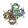
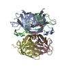
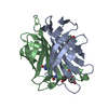
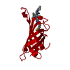
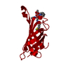
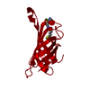
| Title | scdSav(SARK)mv2 - Engineering Single-Chain Dimeric Streptavidin as Host for Artificial Metalloenzymes | |||||||||||||||
 Components Components | Streptavidin | |||||||||||||||
 Keywords Keywords | biotin-binding protein / Artificial Transfer Hydrogenase / Beta Barrel / Streptavidin / TRANSPORT PROTEIN | |||||||||||||||
| Function / homology |  Function and homology information Function and homology information | |||||||||||||||
| Biological species |  Streptomyces avidinii (bacteria) Streptomyces avidinii (bacteria) | |||||||||||||||
| Method | X-RAY DIFFRACTION / SYNCHROTRON / MOLECULAR REPLACEMENT / Resolution: 2 Å | |||||||||||||||
 Authors Authors | Rebelein, J.G. | |||||||||||||||
| Funding support |  Switzerland, Switzerland,  Germany, 4items Germany, 4items
| |||||||||||||||
 Citation Citation | Journal: J.Am.Chem.Soc. / Year: 2019 Title: Breaking Symmetry: Engineering Single-Chain Dimeric Streptavidin as Host for Artificial Metalloenzymes. Authors: Wu, S. / Zhou, Y. / Rebelein, J.G. / Kuhn, M. / Mallin, H. / Zhao, J. / Igareta, N.V. / Ward, T.R. | |||||||||||||||
| History |
|
- Structure visualization
| Structure viewer | Molecule: MolmilJmol/JSmol |
|---|
- Downloads & links
-Download
| PDBx/mmCIF format | 6s50.cif.gz | 119.4 KB | Display | PDBx/mmCIF format |
|---|---|---|---|---|
| PDB format | pdb6s50.ent.gz | 89.8 KB | Display | PDB format |
| PDBx/mmJSON format | 6s50.json.gz | Tree view | PDBx/mmJSON format | |
| Others |  Other downloads Other downloads |
-Validation report
| Arichive directory | https://data.pdbj.org/pub/pdb/validation_reports/s5/6s50ftp://data.pdbj.org/pub/pdb/validation_reports/s5/6s50 | HTTPS FTP |
|---|
-Related structure data
| Related structure data |  6s4qC 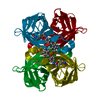 3pk2S S: Starting model for refinement C: citing same article ( |
|---|---|
| Similar structure data |


-Links
 PDBj
PDBj- Assembly
| Deposited unit | 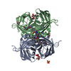
| ||||||||
|---|---|---|---|---|---|---|---|---|---|
| 1 |
| ||||||||
| Unit cell |
|
-Components
| #1: Protein | Mass: 34961.832 Da / Num. of mol.: 2 Source method: isolated from a genetically manipulated source Source: (gene. exp.) Streptomyces avidinii (bacteria) / Production host: Escherichia coli (E. coli) / References: UniProt: P22629#2: Chemical | ChemComp-4IR / {   Mass: 807.488 Da / Num. of mol.: 4 / Source method: obtained synthetically / Formula: C28H45ClIrN5O4S2 Mass: 807.488 Da / Num. of mol.: 4 / Source method: obtained synthetically / Formula: C28H45ClIrN5O4S2#3: Chemical | ChemComp-GOL / Glycerol  Mass: 92.094 Da / Num. of mol.: 6 / Source method: obtained synthetically / Formula: C3H8O3 Mass: 92.094 Da / Num. of mol.: 6 / Source method: obtained synthetically / Formula: C3H8O3#4: Chemical | ChemComp-SO4 / | Sulfate  Mass: 96.063 Da / Num. of mol.: 1 / Source method: obtained synthetically / Formula: SO4 Mass: 96.063 Da / Num. of mol.: 1 / Source method: obtained synthetically / Formula: SO4#5: Water | ChemComp-HOH / | Water Mass: 18.015 Da / Num. of mol.: 129 / Source method: isolated from a natural source / Formula: H2O Mass: 18.015 Da / Num. of mol.: 129 / Source method: isolated from a natural source / Formula: H2OHas ligand of interest | N | |
|---|
-Experimental details
-Experiment
| Experiment | Method: X-RAY DIFFRACTION / Number of used crystals: 1 |
|---|
- Sample preparation
| Crystal | Density Matthews: 2.08 Å3/Da / Density % sol: 40.82 % |
|---|---|
| Crystal grow | Temperature: 293 K / Method: vapor diffusion, sitting drop / pH: 4 / Details: 2 M (NH4)2SO4, 0.1 M Na-Acetate, pH 4 |
-Data collection
| Diffraction | Mean temperature: 100 K / Serial crystal experiment: N |
|---|---|
| Diffraction source | Source: SYNCHROTRON / Site: SLS / Beamline: X06SA / Wavelength: 1 Å |
| Detector | Type: DECTRIS EIGER X 16M / Detector: PIXEL / Date: Sep 30, 2018 |
| Radiation | Protocol: SINGLE WAVELENGTH / Monochromatic (M) / Laue (L): M / Scattering type: x-ray |
| Radiation wavelength | Wavelength: 1 Å / Relative weight: 1 |
| Reflection | Resolution: 2→40.99 Å / Num. obs: 38602 / % possible obs: 99.89 % / Redundancy: 3.3 % / CC1/2: 0.996 / Rmerge(I) obs: 0.0678 / Net I/σ(I): 9.8 |
| Reflection shell | Resolution: 2→2.071 Å / Redundancy: 2.9 % / Rmerge(I) obs: 0.3786 / Mean I/σ(I) obs: 2.49 / Num. unique obs: 3856 / CC1/2: 0.78 / % possible all: 99.79 |
- Processing
| Software |
| |||||||||||||||||||||||||||||||||||||||||||||||||||||||||||||||||||||||||||||||||||||||||||||||||||||||||||||||||||||||||||||||||||||
|---|---|---|---|---|---|---|---|---|---|---|---|---|---|---|---|---|---|---|---|---|---|---|---|---|---|---|---|---|---|---|---|---|---|---|---|---|---|---|---|---|---|---|---|---|---|---|---|---|---|---|---|---|---|---|---|---|---|---|---|---|---|---|---|---|---|---|---|---|---|---|---|---|---|---|---|---|---|---|---|---|---|---|---|---|---|---|---|---|---|---|---|---|---|---|---|---|---|---|---|---|---|---|---|---|---|---|---|---|---|---|---|---|---|---|---|---|---|---|---|---|---|---|---|---|---|---|---|---|---|---|---|---|---|---|
| Refinement | Method to determine structure: MOLECULAR REPLACEMENT Starting model: 3PK2 Resolution: 2→40.99 Å / SU ML: 0.25 / Cross valid method: FREE R-VALUE / σ(F): 1.34 / Phase error: 23.88
| |||||||||||||||||||||||||||||||||||||||||||||||||||||||||||||||||||||||||||||||||||||||||||||||||||||||||||||||||||||||||||||||||||||
| Solvent computation | Shrinkage radii: 0.9 Å / VDW probe radii: 1.11 Å | |||||||||||||||||||||||||||||||||||||||||||||||||||||||||||||||||||||||||||||||||||||||||||||||||||||||||||||||||||||||||||||||||||||
| Refinement step | Cycle: LAST / Resolution: 2→40.99 Å
| |||||||||||||||||||||||||||||||||||||||||||||||||||||||||||||||||||||||||||||||||||||||||||||||||||||||||||||||||||||||||||||||||||||
| Refine LS restraints |
| |||||||||||||||||||||||||||||||||||||||||||||||||||||||||||||||||||||||||||||||||||||||||||||||||||||||||||||||||||||||||||||||||||||
| LS refinement shell |
|
