[English] 日本語
Yorodumi
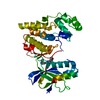
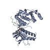
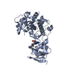
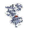
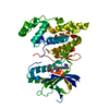
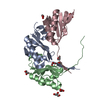
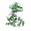
- PDB-4g1w: Crystal structure of JNK1 in complex with JIP1 peptide and 7-Fluo... -

+
Open data


ID or keywords:

Loading...

-
Basic information

Entry
Database: PDB / ID: 4g1w
TitleCrystal structure of JNK1 in complex with JIP1 peptide and 7-Fluoro-3-[4-(2-hydroxy-ethanesulfonyl)-benzyl]-4-oxo-1-phenyl-1,4-dihydro-quinoline-2-carboxylic acid methyl ester
Components
  • C-Jun-amino-terminal kinase-interacting protein 1
  • Mitogen-activated protein kinase 8
KeywordsTransferase/Transferase Inhibitor / kinase inhibitor / Transferase-Transferase Inhibitor complex
Function / homology
Function and homology information


cellular response to stress / positive regulation of cell killing / dentate gyrus mossy fiber / JUN phosphorylation / regulation of CD8-positive, alpha-beta T cell proliferation / regulation of DNA replication origin binding / Interleukin-38 signaling / Activation of BMF and translocation to mitochondria / basal dendrite / Activation of BIM and translocation to mitochondria ...cellular response to stress / positive regulation of cell killing / dentate gyrus mossy fiber / JUN phosphorylation / regulation of CD8-positive, alpha-beta T cell proliferation / regulation of DNA replication origin binding / Interleukin-38 signaling / Activation of BMF and translocation to mitochondria / basal dendrite / Activation of BIM and translocation to mitochondria / JUN kinase activity / WNT5:FZD7-mediated leishmania damping / negative regulation of JUN kinase activity / MAP-kinase scaffold activity / protein serine/threonine kinase binding / JUN kinase binding / positive regulation of cyclase activity / histone deacetylase regulator activity / positive regulation of NLRP3 inflammasome complex assembly / DSCAM interactions / NRAGE signals death through JNK / protein kinase inhibitor activity / Activation of the AP-1 family of transcription factors / Fc-epsilon receptor signaling pathway / kinesin binding / regulation of JNK cascade / MAP kinase activity / regulation of macroautophagy / mitogen-activated protein kinase / negative regulation of intrinsic apoptotic signaling pathway / stress-activated MAPK cascade / response to mechanical stimulus / response to UV / JNK cascade / vesicle-mediated transport / cellular response to cadmium ion / cellular response to amino acid starvation / positive regulation of protein metabolic process / NRIF signals cell death from the nucleus / JNK (c-Jun kinases) phosphorylation and activation mediated by activated human TAK1 / negative regulation of protein binding / mitochondrial membrane / FCERI mediated MAPK activation / positive regulation of JNK cascade / peptidyl-threonine phosphorylation / regulation of circadian rhythm / cellular response to reactive oxygen species / cellular response to mechanical stimulus / histone deacetylase binding / rhythmic process / regulation of protein localization / cellular senescence / Recruitment and ATM-mediated phosphorylation of repair and signaling proteins at DNA double strand breaks / cellular response to oxidative stress / peptidyl-serine phosphorylation / protein phosphatase binding / Oxidative Stress Induced Senescence / response to oxidative stress / cellular response to lipopolysaccharide / positive regulation of apoptotic process / axon / phosphorylation / protein phosphorylation / protein serine kinase activity / protein serine/threonine kinase activity / neuronal cell body / dendrite / synapse / endoplasmic reticulum membrane / positive regulation of gene expression / regulation of DNA-templated transcription / negative regulation of apoptotic process / perinuclear region of cytoplasm / enzyme binding / nucleoplasm / ATP binding / nucleus / plasma membrane / cytosol / cytoplasm
Similarity search - Function
JIP1, SH3 domain / : / Mitogen-activated protein (MAP) kinase, JNK / Phosphotyrosine interaction domain (PTB/PID) / Phosphotyrosine interaction domain (PID) profile. / Phosphotyrosine-binding domain, phosphotyrosine-interaction (PI) domain / PTB/PI domain / Variant SH3 domain / Mitogen-activated protein (MAP) kinase, conserved site / MAP kinase signature. ...JIP1, SH3 domain / : / Mitogen-activated protein (MAP) kinase, JNK / Phosphotyrosine interaction domain (PTB/PID) / Phosphotyrosine interaction domain (PID) profile. / Phosphotyrosine-binding domain, phosphotyrosine-interaction (PI) domain / PTB/PI domain / Variant SH3 domain / Mitogen-activated protein (MAP) kinase, conserved site / MAP kinase signature. / Src homology 3 domains / SH3-like domain superfamily / Src homology 3 (SH3) domain profile. / SH3 domain / PH-like domain superfamily / Transferase(Phosphotransferase) domain 1 / Transferase(Phosphotransferase); domain 1 / Phosphorylase Kinase; domain 1 / Phosphorylase Kinase; domain 1 / Serine/threonine-protein kinase, active site / Serine/Threonine protein kinases active-site signature. / Protein kinase domain / Serine/Threonine protein kinases, catalytic domain / Protein kinase domain profile. / Protein kinase domain / Protein kinase-like domain superfamily / 2-Layer Sandwich / Orthogonal Bundle / Mainly Alpha / Alpha Beta
Similarity search - Domain/homology
Chem-G1W / Stress-activated protein kinase JNK / Mitogen-activated protein kinase 8 / C-Jun-amino-terminal kinase-interacting protein 1
Similarity search - Component
Biological speciesHomo sapiens (human)
MethodX-RAY DIFFRACTION / SYNCHROTRON / FOURIER SYNTHESIS / Resolution: 2.45 Å
AuthorsKuglstatter, A. / Shao, A.
CitationJournal: To be Published
Title: Crystal structure of JNK1 in complex with JIP1 peptide and inhibitor
Authors: Gong, L. / Tan, Y.-C. / Boice, G. / Abbot, S.
History
DepositionJul 11, 2012Deposition site: RCSB / Processing site: RCSB
Revision 1.0Jul 17, 2013Provider: repository / Type: Initial release
Revision 1.1Sep 13, 2023Group: Data collection / Database references ...Data collection / Database references / Derived calculations / Refinement description
Category: chem_comp_atom / chem_comp_bond ...chem_comp_atom / chem_comp_bond / database_2 / pdbx_initial_refinement_model / struct_ref_seq_dif / struct_site
Item: _database_2.pdbx_DOI / _database_2.pdbx_database_accession ..._database_2.pdbx_DOI / _database_2.pdbx_database_accession / _struct_ref_seq_dif.details / _struct_site.pdbx_auth_asym_id / _struct_site.pdbx_auth_comp_id / _struct_site.pdbx_auth_seq_id

-
Structure visualization

Structure viewerMolecule:
MolmilJmol/JSmol

Downloads & links

-
Assembly

Deposited unit
A: Mitogen-activated protein kinase 8
B: C-Jun-amino-terminal kinase-interacting protein 1
hetero molecules


Theoretical massNumber of molelcules
Total (without water)44,4293
Polymers43,9342
Non-polymers4961
Water77543
1


  • Idetical with deposited unit
  • defined by software
TypeNameSymmetry operationNumber
identity operation1_555x,y,z1
Buried area1200 Å2
ΔGint-8 kcal/mol
Surface area16150 Å2
MethodPISA
Unit cell
Length a, b, c (Å)62.008, 80.308, 85.102
Angle α, β, γ (deg.)90.00, 90.00, 90.00
Int Tables number19
Space group name H-MP212121
DetailsAS PER AUTHORS JNK1 IS PRESENT AS A MONOMER IN BIOLOGICAL ASSEMBLY

-
Components

#1: Protein Mitogen-activated protein kinase 8 / Mitogen-activated protein kinase 8 isoform JNK1 beta2 / Mitogen-activated protein kinase 8 / ...Mitogen-activated protein kinase 8 isoform JNK1 beta2 / Mitogen-activated protein kinase 8 / isoform CRA_d / cDNA FLJ77387 / highly similar to Homo sapiens mitogen-activated protein kinase 8 (MAPK8) / transcript variant 4 / mRNA


Mass: 42588.191 Da / Num. of mol.: 1 / Fragment: RESIDUES 1-363
Source method: isolated from a genetically manipulated source
Source: (gene. exp.) Homo sapiens (human) / Gene: MAPK8, hCG_23734 / Production host: Escherichia coli (E. coli)
References: UniProt: A1L4K2, UniProt: P45983*PLUS, EC: 2.7.1.37
#2: Protein/peptide C-Jun-amino-terminal kinase-interacting protein 1 / JIP-1 / JNK-interacting protein 1 / Islet-brain 1 / IB-1 / JNK MAP kinase scaffold protein 1 / ...JIP-1 / JNK-interacting protein 1 / Islet-brain 1 / IB-1 / JNK MAP kinase scaffold protein 1 / Mitogen-activated protein kinase 8-interacting protein 1


Mass: 1345.612 Da / Num. of mol.: 1 / Fragment: RESIDUES 157-167 / Source method: obtained synthetically / Source: (synth.) Homo sapiens (human) / References: UniProt: Q9UQF2
#3: Chemical ChemComp-G1W / methyl 7-fluoro-3-{4-[(2-hydroxyethyl)sulfonyl]benzyl}-4-oxo-1-phenyl-1,4-dihydroquinoline-2-carboxylate


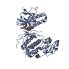
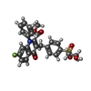
Mass: 495.519 Da / Num. of mol.: 1 / Source method: obtained synthetically / Formula: C26H22FNO6S
#4: Water ChemComp-HOH / water / Water


Mass: 18.015 Da / Num. of mol.: 43 / Source method: isolated from a natural source / Formula: H2O

-
Experimental details

-
Experiment

ExperimentMethod: X-RAY DIFFRACTION / Number of used crystals: 1

-
Sample preparation

CrystalDensity Matthews: 2.41 Å3/Da / Density % sol: 48.99 %
Crystal growTemperature: 289 K / Method: vapor diffusion, hanging drop / pH: 5.5
Details: 20% PEG 3350, 0.2 M LiSO4, 0.1 M BIS-TRIS, pH 5.5, VAPOR DIFFUSION, HANGING DROP, temperature 289K

-
Data collection

DiffractionMean temperature: 100 K
Diffraction sourceSource: SYNCHROTRON / Site: ALS / Beamline: 5.0.1 / Wavelength: 1 Å
DetectorType: ADSC QUANTUM 210 / Detector: CCD
RadiationProtocol: SINGLE WAVELENGTH / Monochromatic (M) / Laue (L): M / Scattering type: x-ray
Radiation wavelengthWavelength: 1 Å / Relative weight: 1
ReflectionResolution: 2.45→50 Å / Num. obs: 15444 / % possible obs: 95.3 % / Redundancy: 5.3 % / Biso Wilson estimate: 67.5 Å2 / Rsym value: 0.043 / Net I/σ(I): 24.8
Reflection shellResolution: 2.45→2.54 Å / Redundancy: 4.6 % / Mean I/σ(I) obs: 2.5 / Num. unique all: 1451 / Rsym value: 0.47 / % possible all: 92.6

-
Processing

Software
NameVersionClassification
ADSCQuantumdata collection
MOLREPphasing
REFMAC5.2.0019refinement
DENZOdata reduction
SCALEPACKdata scaling
RefinementMethod to determine structure: FOURIER SYNTHESIS
Starting model: PDB entry 1UKH
Resolution: 2.45→37.6 Å / Cor.coef. Fo:Fc: 0.942 / Cor.coef. Fo:Fc free: 0.893 / SU B: 24.294 / SU ML: 0.248 / Cross valid method: THROUGHOUT / ESU R: 0.509 / ESU R Free: 0.326 / Stereochemistry target values: MAXIMUM LIKELIHOOD
RfactorNum. reflection% reflectionSelection details
Rfree0.29689 771 5 %RANDOM
Rwork0.23027 ---
obs0.2334 14642 95.34 %-
Solvent computationIon probe radii: 0.8 Å / Shrinkage radii: 0.8 Å / VDW probe radii: 1.2 Å / Solvent model: BABINET MODEL WITH MASK
Displacement parametersBiso mean: 75.213 Å2
Baniso -1Baniso -2Baniso -3
1-0.81 Å20 Å20 Å2
2---0.46 Å20 Å2
3----0.35 Å2
Refinement stepCycle: LAST / Resolution: 2.45→37.6 Å
ProteinNucleic acidLigandSolventTotal
Num. atoms2674 0 35 43 2752
Refine LS restraints
Refine-IDTypeDev idealDev ideal targetNumber
X-RAY DIFFRACTIONr_bond_refined_d0.0120.0222774
X-RAY DIFFRACTIONr_bond_other_d
X-RAY DIFFRACTIONr_angle_refined_deg1.261.9913753
X-RAY DIFFRACTIONr_angle_other_deg
X-RAY DIFFRACTIONr_dihedral_angle_1_deg5.0345326
X-RAY DIFFRACTIONr_dihedral_angle_2_deg40.02324.309123
X-RAY DIFFRACTIONr_dihedral_angle_3_deg15.47615504
X-RAY DIFFRACTIONr_dihedral_angle_4_deg18.141515
X-RAY DIFFRACTIONr_chiral_restr0.0840.2410
X-RAY DIFFRACTIONr_gen_planes_refined0.0040.022082
X-RAY DIFFRACTIONr_gen_planes_other
X-RAY DIFFRACTIONr_nbd_refined0.2130.21147
X-RAY DIFFRACTIONr_nbd_other
X-RAY DIFFRACTIONr_nbtor_refined0.3050.21882
X-RAY DIFFRACTIONr_nbtor_other
X-RAY DIFFRACTIONr_xyhbond_nbd_refined0.1460.274
X-RAY DIFFRACTIONr_xyhbond_nbd_other
X-RAY DIFFRACTIONr_metal_ion_refined
X-RAY DIFFRACTIONr_metal_ion_other
X-RAY DIFFRACTIONr_symmetry_vdw_refined0.2340.217
X-RAY DIFFRACTIONr_symmetry_vdw_other
X-RAY DIFFRACTIONr_symmetry_hbond_refined0.2270.23
X-RAY DIFFRACTIONr_symmetry_hbond_other
X-RAY DIFFRACTIONr_symmetry_metal_ion_refined
X-RAY DIFFRACTIONr_symmetry_metal_ion_other
X-RAY DIFFRACTIONr_mcbond_it1.0011.51704
X-RAY DIFFRACTIONr_mcbond_other
X-RAY DIFFRACTIONr_mcangle_it1.48722690
X-RAY DIFFRACTIONr_scbond_it1.82131221
X-RAY DIFFRACTIONr_scangle_it2.8084.51063
X-RAY DIFFRACTIONr_rigid_bond_restr
X-RAY DIFFRACTIONr_sphericity_free
X-RAY DIFFRACTIONr_sphericity_bonded
LS refinement shellResolution: 2.45→2.514 Å / Total num. of bins used: 20
RfactorNum. reflection% reflection
Rfree0.374 50 -
Rwork0.319 1003 -
obs--90.7 %
Refinement TLS params.Method: refined / Origin x: -5.7753 Å / Origin y: 8.6916 Å / Origin z: -20.3257 Å
111213212223313233
T0.0378 Å2-0.0521 Å2-0.0022 Å2-0.0304 Å2-0.0265 Å2---0.0103 Å2
L1.4873 °2-0.0876 °20.3461 °2-0.9774 °2-0.2312 °2--0.1688 °2
S-0.0083 Å °-0.019 Å °0.0596 Å °0.0482 Å °0.0175 Å °0.141 Å °0.0242 Å °0.0259 Å °-0.0092 Å °
Refinement TLS group
IDRefine-IDRefine TLS-IDAuth asym-IDAuth seq-ID
1X-RAY DIFFRACTION1A501 - 543
2X-RAY DIFFRACTION1A401
3X-RAY DIFFRACTION1B154 - 162
4X-RAY DIFFRACTION1A8 - 361

+
About Yorodumi

-
News

-
Feb 9, 2022. New format data for meta-information of EMDB entries

New format data for meta-information of EMDB entries

  • Version 3 of the EMDB header file is now the official format.
  • The previous official version 1.9 will be removed from the archive.

Related info.:EMDB header

External links:wwPDB to switch to version 3 of the EMDB data model

-
Aug 12, 2020. Covid-19 info

Covid-19 info

URL: https://pdbj.org/emnavi/covid19.php

New page: Covid-19 featured information page in EM Navigator.

Related info.:Covid-19 info / Mar 5, 2020. Novel coronavirus structure data

+
Mar 5, 2020. Novel coronavirus structure data

Novel coronavirus structure data

Related info.:Yorodumi Speices / Aug 12, 2020. Covid-19 info

External links:COVID-19 featured content - PDBj / Molecule of the Month (242):Coronavirus Proteases

+
Jan 31, 2019. EMDB accession codes are about to change! (news from PDBe EMDB page)

EMDB accession codes are about to change! (news from PDBe EMDB page)

  • The allocation of 4 digits for EMDB accession codes will soon come to an end. Whilst these codes will remain in use, new EMDB accession codes will include an additional digit and will expand incrementally as the available range of codes is exhausted. The current 4-digit format prefixed with “EMD-” (i.e. EMD-XXXX) will advance to a 5-digit format (i.e. EMD-XXXXX), and so on. It is currently estimated that the 4-digit codes will be depleted around Spring 2019, at which point the 5-digit format will come into force.
  • The EM Navigator/Yorodumi systems omit the EMD- prefix.

Related info.:Q: What is EMD? / ID/Accession-code notation in Yorodumi/EM Navigator

External links:EMDB Accession Codes are Changing Soon! / Contact to PDBj

+
Jul 12, 2017. Major update of PDB

Major update of PDB

  • wwPDB released updated PDB data conforming to the new PDBx/mmCIF dictionary.
  • This is a major update changing the version number from 4 to 5, and with Remediation, in which all the entries are updated.
  • In this update, many items about electron microscopy experimental information are reorganized (e.g. em_software).
  • Now, EM Navigator and Yorodumi are based on the updated data.

External links:wwPDB Remediation / Enriched Model Files Conforming to OneDep Data Standards Now Available in the PDB FTP Archive

-
Yorodumi

Thousand views of thousand structures

  • Yorodumi is a browser for structure data from EMDB, PDB, SASBDB, etc.
  • This page is also the successor to EM Navigator detail page, and also detail information page/front-end page for Omokage search.
  • The word "yorodu" (or yorozu) is an old Japanese word meaning "ten thousand". "mi" (miru) is to see.

Related info.:EMDB / PDB / SASBDB / Comparison of 3 databanks / Yorodumi Search / Aug 31, 2016. New EM Navigator & Yorodumi / Yorodumi Papers / Jmol/JSmol / Function and homology information / Changes in new EM Navigator and Yorodumi

Read more