 Movie
Movie Controller

[English] 日本語
 Yorodumi
- PDB-4u79: Crystal structure of human JNK3 in complex with a benzenesulfonam... -
+ Open data

- Basic information
| Entry | Database: PDB / ID: 4u79 | ||||||
|---|---|---|---|---|---|---|---|
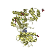
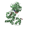
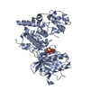
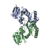
| Title | Crystal structure of human JNK3 in complex with a benzenesulfonamide inhibitor. | ||||||
 Components Components | Mitogen-activated protein kinase 10 | ||||||
 Keywords Keywords | TRANSFERASE/TRANSFERASE INHIBITOR /  Transferase / TRANSFERASE-TRANSFERASE INHIBITOR complex Transferase / TRANSFERASE-TRANSFERASE INHIBITOR complex | ||||||
| Function / homology |  Function and homology information Function and homology informationJUN kinase activity / Activation of the AP-1 family of transcription factors / Fc-epsilon receptor signaling pathway / MAP kinase kinase activity / response to light stimulus / mitogen-activated protein kinase / JNK cascade / JNK (c-Jun kinases) phosphorylation and activation mediated by activated human TAK1 / FCERI mediated MAPK activation / regulation of circadian rhythm ...JUN kinase activity / Activation of the AP-1 family of transcription factors / Fc-epsilon receptor signaling pathway / MAP kinase kinase activity / response to light stimulus / mitogen-activated protein kinase / JNK cascade / JNK (c-Jun kinases) phosphorylation and activation mediated by activated human TAK1 / FCERI mediated MAPK activation / regulation of circadian rhythm / cellular senescence / rhythmic process / Oxidative Stress Induced Senescence / protein phosphorylation / protein serine kinase activity / signal transduction / mitochondrion / nucleoplasm / ATP binding / nucleus / plasma membrane / cytosol / cytoplasmSimilarity search - Function | ||||||
| Biological species |  Homo sapiens (human) Homo sapiens (human) | ||||||
| Method | X-RAY DIFFRACTION / SYNCHROTRON / MOLECULAR REPLACEMENT / Resolution: 2.23 Å | ||||||
 Authors Authors | Mohr, C. | ||||||
 Citation Citation | Journal: Acs Med.Chem.Lett. / Year: 2015 Title: Unfolded Protein Response in Cancer: IRE1 alpha Inhibition by Selective Kinase Ligands Does Not Impair Tumor Cell Viability. Authors: Harrington, P.E. / Biswas, K. / Malwitz, D. / Tasker, A.S. / Mohr, C. / Andrews, K.L. / Dellamaggiore, K. / Kendall, R. / Beckmann, H. / Jaeckel, P. / Materna-Reichelt, S. / Allen, J.R. / Lipford, J.R. | ||||||
| History |
|
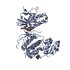
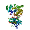
- Structure visualization
| Structure viewer | Molecule: MolmilJmol/JSmol |
|---|
- Downloads & links
-Download
| PDBx/mmCIF format | 4u79.cif.gz | 152.9 KB | Display | PDBx/mmCIF format |
|---|---|---|---|---|
| PDB format | pdb4u79.ent.gz | 119 KB | Display | PDB format |
| PDBx/mmJSON format | 4u79.json.gz | Tree view | PDBx/mmJSON format | |
| Others |  Other downloads Other downloads |
-Validation report
| Arichive directory | https://data.pdbj.org/pub/pdb/validation_reports/u7/4u79ftp://data.pdbj.org/pub/pdb/validation_reports/u7/4u79 | HTTPS FTP |
|---|
-Related structure data
| Related structure data |  4u6rC  3da6S C: citing same article ( S: Starting model for refinement |
|---|---|
| Similar structure data |


-Links
 PDBj
PDBj


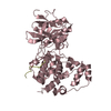
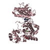
- Assembly
| Deposited unit | 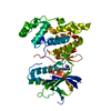
| ||||||||
|---|---|---|---|---|---|---|---|---|---|
| 1 |
| ||||||||
| Unit cell |
|
-Components
| #1: Protein | Mass: 42059.676 Da / Num. of mol.: 1 / Fragment: UNP residues 39-402 Source method: isolated from a genetically manipulated source Source: (gene. exp.) Homo sapiens (human) / Gene: MAPK10, JNK3, JNK3A, PRKM10, SAPK1B / Production host:  Escherichia coli (E. coli) Escherichia coli (E. coli)References: UniProt: P53779, mitogen-activated protein kinase |
|---|---|
| #2: Chemical | ChemComp-3EL /   Mass: 566.673 Da / Num. of mol.: 1 / Source method: obtained synthetically / Formula: C31H30N6O3S Mass: 566.673 Da / Num. of mol.: 1 / Source method: obtained synthetically / Formula: C31H30N6O3S |
| #3: Water | ChemComp-HOH / Water Mass: 18.015 Da / Num. of mol.: 33 / Source method: isolated from a natural source / Formula: H2O Mass: 18.015 Da / Num. of mol.: 33 / Source method: isolated from a natural source / Formula: H2O |
-Experimental details
-Experiment
| Experiment | Method: X-RAY DIFFRACTION |
|---|
- Sample preparation
| Crystal | Density Matthews: 2.49 Å3/Da / Density % sol: 50.67 % |
|---|---|
| Crystal grow | Temperature: 277 K / Method: vapor diffusion, hanging drop / pH: 7.5 / Details: 0.1M HEPES, 20mMBMe, 30%PEG550MME, pH7.5 |
-Data collection
| Diffraction | Mean temperature: 100 K |
|---|---|
| Diffraction source | Source: SYNCHROTRON / Site: APS  / Beamline: 22-ID / Wavelength: 1 Å / Beamline: 22-ID / Wavelength: 1 Å |
| Detector | Type: MARMOSAIC 300 mm CCD / Detector: CCD / Date: Jul 30, 2011 |
| Radiation | Monochromator: double crystal / Protocol: SINGLE WAVELENGTH / Monochromatic (M) / Laue (L): M / Scattering type: x-ray |
| Radiation wavelength | Wavelength: 1 Å / Relative weight: 1 |
| Reflection | Resolution: 2.23→50 Å / Num. obs: 21261 / % possible obs: 99.9 % / Redundancy: 5.8 % / Rmerge(I) obs: 0.127 / Net I/σ(I): 12.18 |
| Reflection shell | Resolution: 2.23→2.3 Å / Redundancy: 5.3 % / Rmerge(I) obs: 0.558 / Mean I/σ(I) obs: 2.04 / % possible all: 99.9 |
- Processing
| Software | Name: REFMAC / Version: 5.8.0071 / Classification: refinement | ||||||||||||||||||||||||||||||||||||||||||||||||||||||||||||||||||||||||||||||||||||||||||||||||||||||||||||||||||||||||||||||||||||||||||||||||||||||||||||||||||||||||||||||||||||||
|---|---|---|---|---|---|---|---|---|---|---|---|---|---|---|---|---|---|---|---|---|---|---|---|---|---|---|---|---|---|---|---|---|---|---|---|---|---|---|---|---|---|---|---|---|---|---|---|---|---|---|---|---|---|---|---|---|---|---|---|---|---|---|---|---|---|---|---|---|---|---|---|---|---|---|---|---|---|---|---|---|---|---|---|---|---|---|---|---|---|---|---|---|---|---|---|---|---|---|---|---|---|---|---|---|---|---|---|---|---|---|---|---|---|---|---|---|---|---|---|---|---|---|---|---|---|---|---|---|---|---|---|---|---|---|---|---|---|---|---|---|---|---|---|---|---|---|---|---|---|---|---|---|---|---|---|---|---|---|---|---|---|---|---|---|---|---|---|---|---|---|---|---|---|---|---|---|---|---|---|---|---|---|---|
| Refinement | Method to determine structure: MOLECULAR REPLACEMENT Starting model: 3DA6 Resolution: 2.23→50 Å / Cor.coef. Fo:Fc: 0.952 / Cor.coef. Fo:Fc free: 0.94 / SU B: 19.089 / SU ML: 0.214 / Cross valid method: THROUGHOUT / ESU R: 0.278 / ESU R Free: 0.212 / Stereochemistry target values: MAXIMUM LIKELIHOOD / Details: HYDROGENS HAVE BEEN ADDED IN THE RIDING POSITIONS
| ||||||||||||||||||||||||||||||||||||||||||||||||||||||||||||||||||||||||||||||||||||||||||||||||||||||||||||||||||||||||||||||||||||||||||||||||||||||||||||||||||||||||||||||||||||||
| Solvent computation | Ion probe radii: 0.8 Å / Shrinkage radii: 0.8 Å / VDW probe radii: 1.2 Å / Solvent model: MASK | ||||||||||||||||||||||||||||||||||||||||||||||||||||||||||||||||||||||||||||||||||||||||||||||||||||||||||||||||||||||||||||||||||||||||||||||||||||||||||||||||||||||||||||||||||||||
| Displacement parameters | Biso mean: 55.958 Å2
| ||||||||||||||||||||||||||||||||||||||||||||||||||||||||||||||||||||||||||||||||||||||||||||||||||||||||||||||||||||||||||||||||||||||||||||||||||||||||||||||||||||||||||||||||||||||
| Refinement step | Cycle: 1 / Resolution: 2.23→50 Å
| ||||||||||||||||||||||||||||||||||||||||||||||||||||||||||||||||||||||||||||||||||||||||||||||||||||||||||||||||||||||||||||||||||||||||||||||||||||||||||||||||||||||||||||||||||||||
| Refine LS restraints |
|